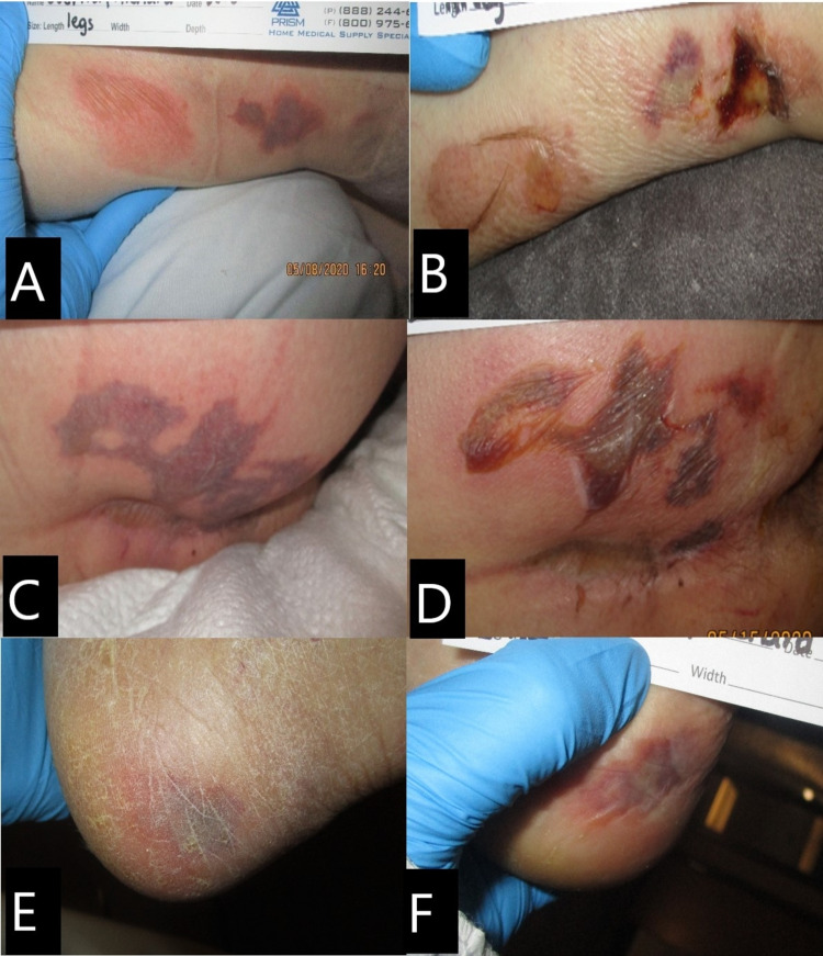
Figure 2. Case 2. Skin lesions.
(A) Right lower leg on day 10: purpuric nonblanchable patch. (B) Right lower leg on day 14: serous-filled bullae with some necrotic tissue. (C) Right buttock on day 10: purpuric nonblanchable patch resembling retiform purpura. (D) Right buttock on day 14: lesion evolved to a serosanguineous-filled bullae (E) Left lateral heel on day 10: purpuric nonblanchable patch. (F) Left lateral heel on day 14: purpuric nonblanchable patch.

