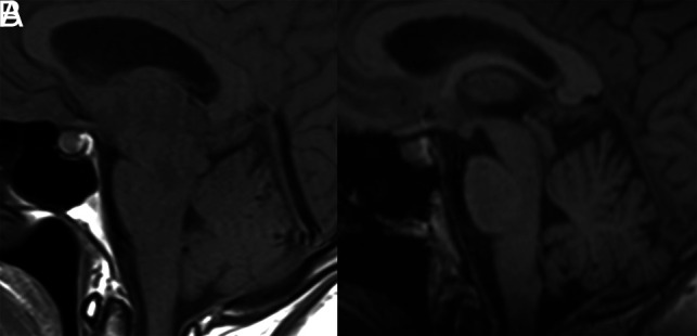
FIG 3.

Pre- and posttreatment MRIs of the patient in Fig 2. A, Sagittal T1-weighted MR imaging shows brain sag with narrowing of the mamillopontine distance, narrowing of the prepontine cistern, and inferior sloping of the floor of the third ventricle. There is also pituitary enlargement and distension of the straight sinus. B, Resolution of these changes following CT-guided injection of fibrin sealant.
