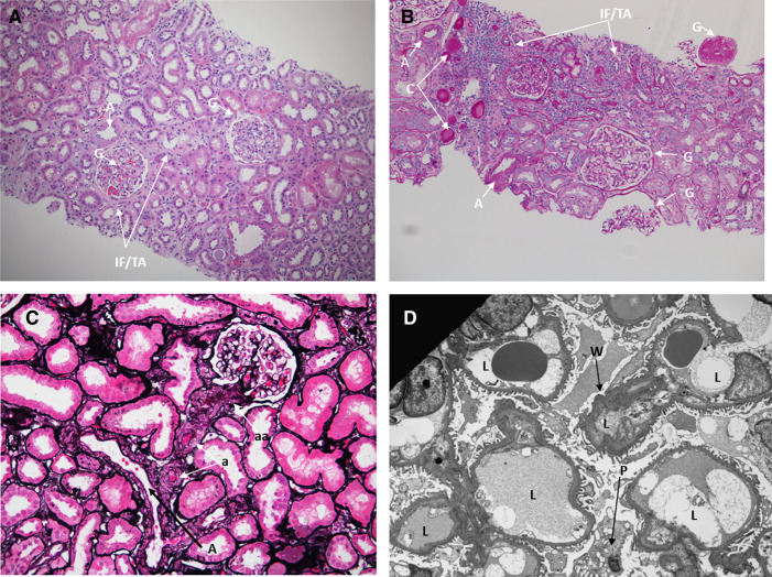
Figure 2.
Pathology assessment of the kidney biopsy specimen. (A) A hematoxylin and eosin-stained section (×100) reveals kidney cortex with glomeruli with normal cellularity (arrows) and focal interstitial fibrosis and tubular atrophy (IF/TA; arrows). An intralobar artery (A) shows mild fibrous thickening. G, global sclerosis. (B) On a periodic acid–Schiff-stained level (×100), there are three nonsclerotic glomeruli, and none have global sclerosis (G). There is focal IF/TA and mild chronic interstitial inflammation. Two arteries (A) within normal limits and proteinaceous casts (C) are also identified. (C) Silver–Jones (×200) shows an artery (A), several arterioles (a) with mild fibrous thickening, and likely afferent arterioles (aa). (D) Electron microscopy (×2550) reveals glomerular capillary loops (L) with mild segmental ischemic wrinkling (W) of the glomerular basement membrane and a vacuolated podocyte (P).

