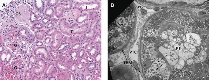
Figure 3.
Pathologic evidence of chronic active kidney cell injury. (A) A hematoxylin and eosin-stained section (×200) shows two normal glomeruli (G) and one glomerulus with global sclerosis (GS). Tubules (T) show several mild changes of acute tubular injury, such as apical blebbing, cytoplasmic vacuoles, and epithelial simplification. Epithelial simplification refers to the loss of columnar architecture and brush borders of the proximal tubular cells and their transition to a flat, squamoid undifferentiated appearance with loss of columnar and brush border. (B) Electron microscopy (×1050) depicts proximal tubule (PT) with normal apical brush border as well as focal epithelial cytoplasmic vacuoles (arrows) and mild thickening of the basement membrane (TBM). Two peritubular capillaries (PTC) are within normal limits.

