Figure 1.

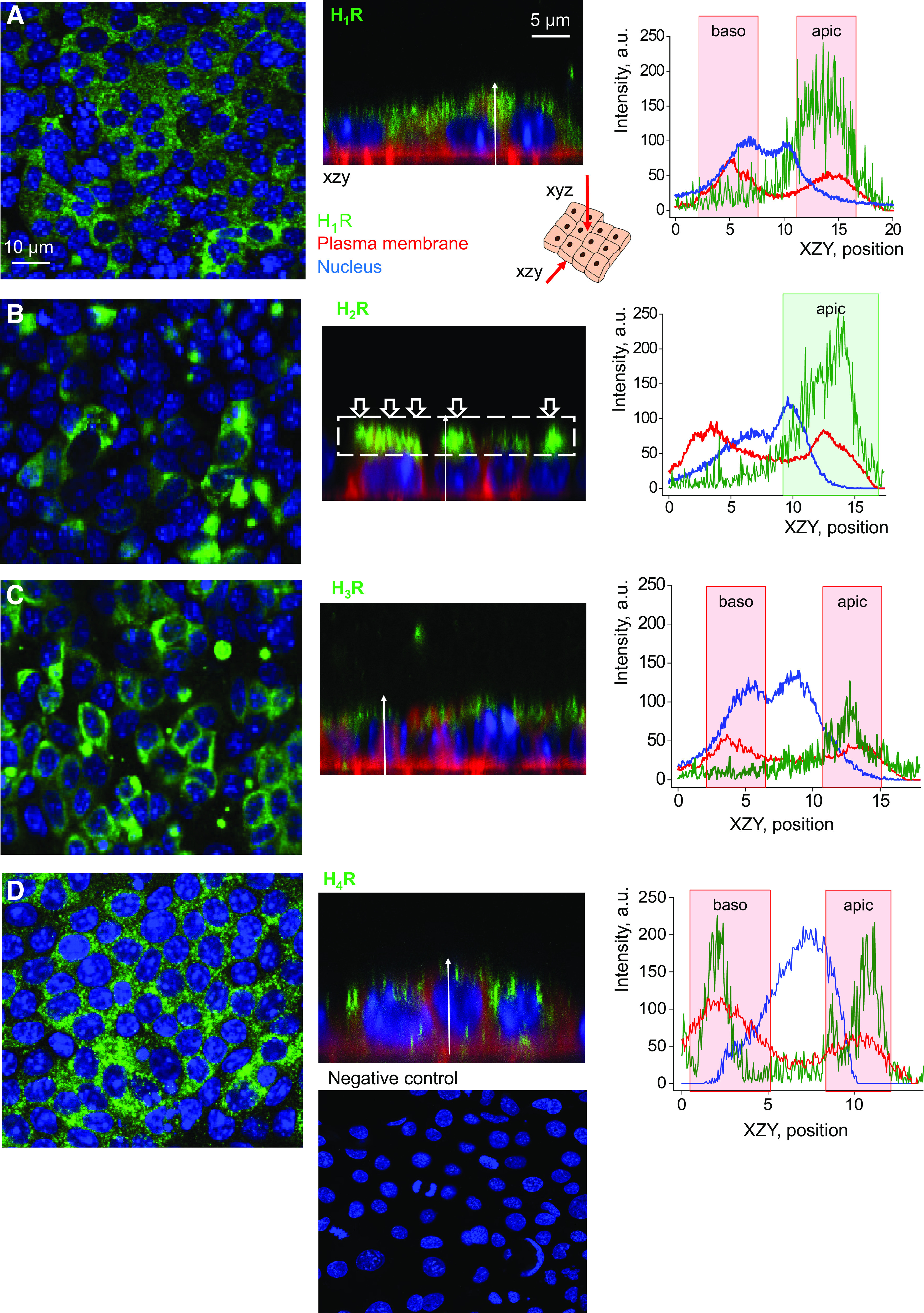
Histamine receptors are detectable in the basolateral and apical membrane of mpkCCDcl4 cells. Shown is the top-down (xyz) and side (xzy) view of immunocytochemistry performed to visualize histamine receptors’ expression [H1R (A), H2R (B), H3R (C), and H4R (D)] in mpkCCDcl4 cells. Scale bar is shown in A, and is the same for all images. Graphs demonstrate representative quantification of fluorescence intensity distribution of the receptors’ level (green), plasma membrane localization (red), and the nucleus (blue) in the xzy view along the white arrow. Baso, basolateral membrane; apic, apical membrane. Shown are representative images, n = 3–5 total independent staining experiments. Bottom panel shows negative control (secondary antibodies in the absence of primary antibodies and Hoechst stain). mpkCCDcl4, mouse cortical collecting duct cell line.
