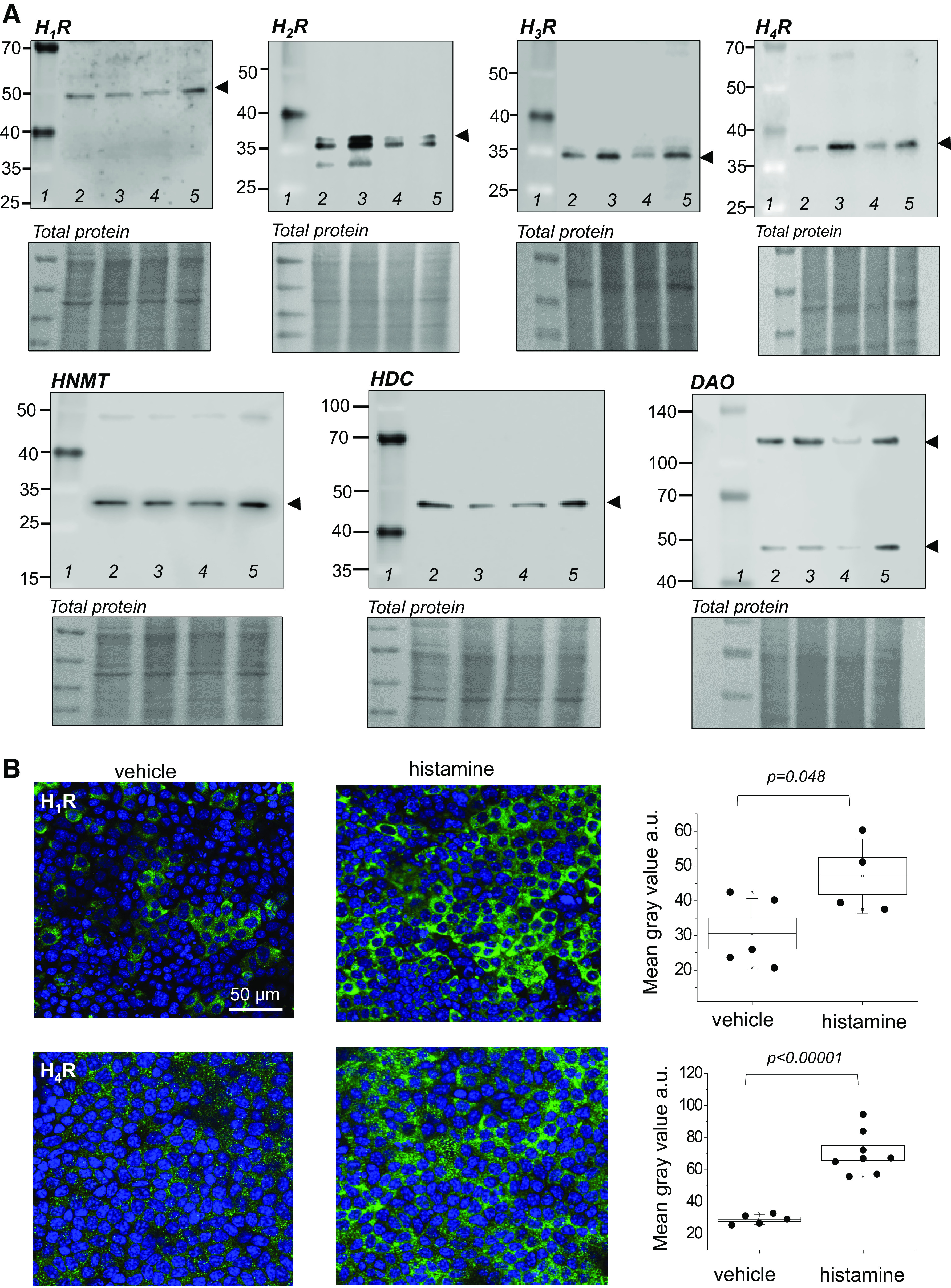
Figure 2.

The components of the histaminergic system and all histamine receptors are present in the collecting duct cells. A: Western blot analysis showing H1R, H2R, H3R, H4R, histamine-N-methyltransferase (HNMT), histidine decarboxylase (HDC), and diamine oxidase (DAO) expression in mpkCCDcl4 cells. Each lane on the Western blot image pertains to an independently grown cell culture dish to illustrate that proteins are consistently expressed in independent cell samples; no treatments were performed. Lane 1 in each Western is a weight marker; lanes 2–5 are individual independent lysates. B: representative immunofluorescence staining of H1R and H4R expression in the apical membrane of the mpkCCDcl4 cells following treatment with vehicle or 200 μM histamine for 4 h; shown are topside view images (XYZ) at ×63. Graphs show quantification of H1R and H4R expression (based on fluorescence intensity, normalized to background fluorescence) between treatment and experimental groups. n = 3 experiments from independent cell samples, n = 4–8 fields of view were quantified. Box illustrated means ± SE, whiskers are SD. Two-sample Student’s t test was used to test for statistical significance in B. Scale bar is shown in B for H1R and is the same for all images. mpkCCDcl4, mouse cortical collecting duct cell line.
