
Figure 3.
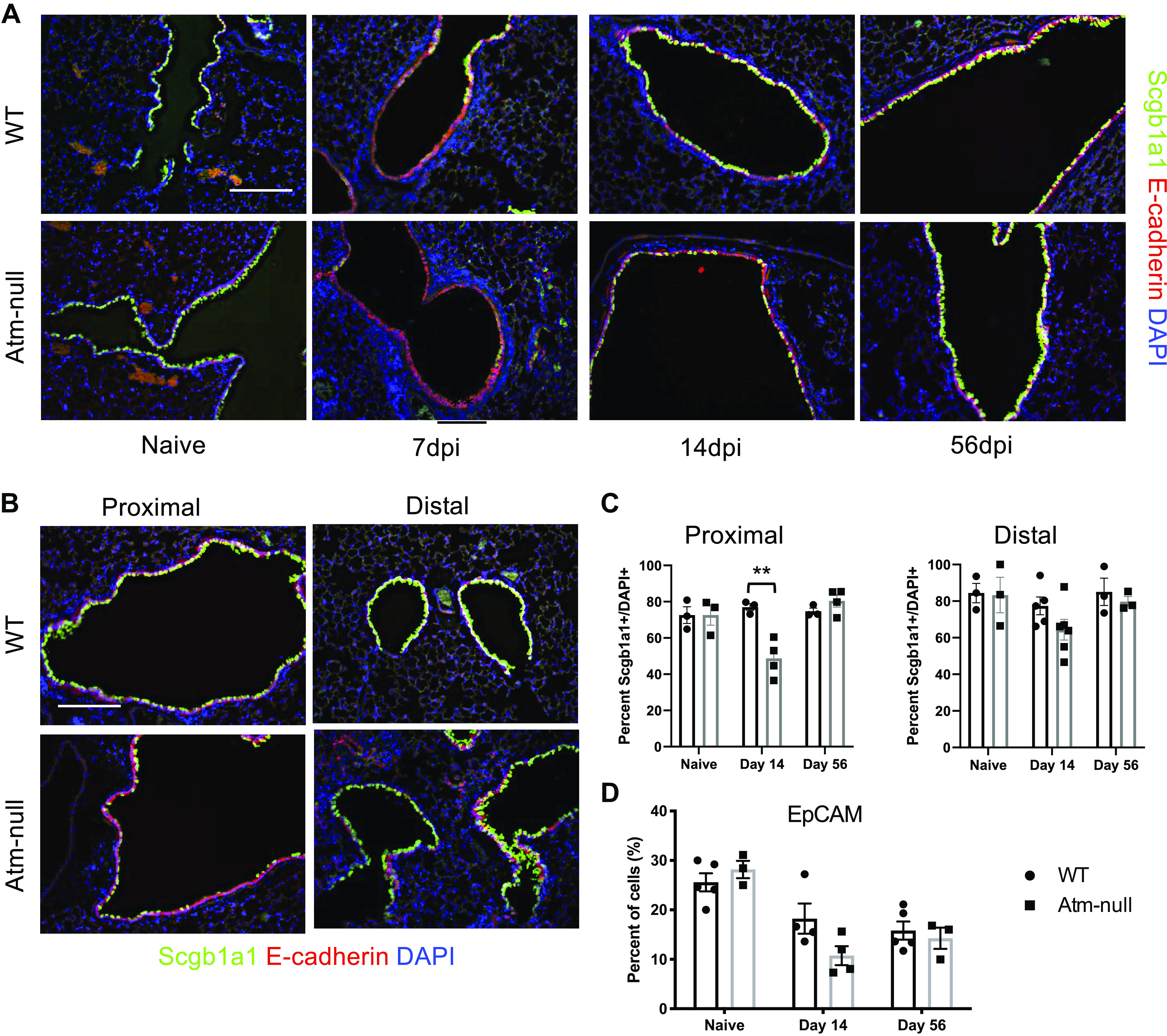
Atm-null mice have aberrant expression of Scgb1a1 following IAV infection. WT and Atm-null mice were infected with 105 PFU of HKx31. Lungs were harvested from naïve mice and at 7, 14, and 56 dpi. A: sections of lung were stained with antibodies against Scgb1a1 (green) and epithelial cadherin (E-cadherin; red), and counterstained with DAPI (blue). B: representative images of proximal and distal airways of WT and Atm-null mice collected at 14 dpi and stained as in A. C: the number of Scgb1a1+ cells in A were counted and graphed as a percentage of the total number of DAPI+ cells per airway. Proximal airways were determined to be larger airways within a section, whereas distal airways are those that contain BADJ. D: whole lung homogenates were stained for EpCAM and run on a flow cytometer. EpCAM+ cells were graphed as a percentage of total cells analyzed. Scale bars = 200 µm. Representative images of n ≥ 3 mice/time point/group are shown. P values were determined by Student’s t test. **P ≤ 0.01; n ≥ 3/time point/group. BADJ, bronchoalveolar duct junction; dpi, days postinfection; EpCAM, epithelial cell adhesion molecule; HKx31, Hong Kong/X31; IAV, influenza A virus; PFU, plaque-forming unit; Scgb1a1, secretoglobin family 1 A member 1; WT, wildtype.