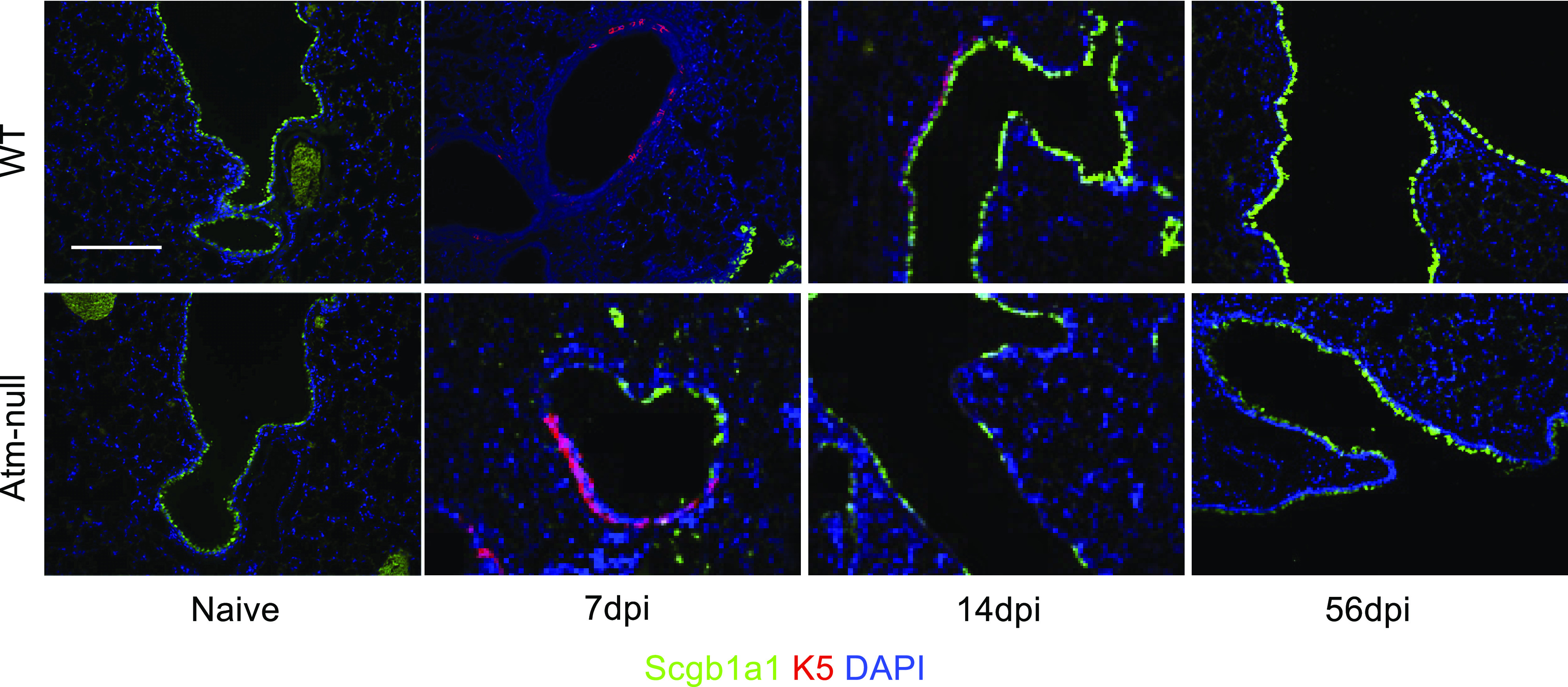
Figure 6.

Atm is not required for K5+ cells to expand into the distal airways following IAV infection. Lungs from naïve and HKx31 infected mice at 7, 14, and 56 dpi were stained with Scgb1a1 (green), cytokeratin 5 (K5; red) and counterstained with DAPI (blue). Images are representative data from n ≥ 3 mice/time point/group. Scale bar = 200 µm. Representative images of n ≥ 3 mice/time point/group are shown. dpi, days postinfection; HKx31, Hong Kong/X31; IAV, influenza A virus; Scgb1a1, secretoglobin family 1 A member 1.
