

Abstract
Background
Studies have shown the promising prospects of rosmarinic acid (RosA) for the prevention and treatment of allergic diseases.
Objective
The aim of this study was to investigate the effects of RosA on inflammatory reaction in rat models of allergic rhinitis (AR) after PM2.5 exposure.
Methods
Allergic rhinitis rat models were established by ovalbumin sensitization, and PM2.5 was applied at a concentration of 1000 μg/m3, 3 h a day for 30 consecutive days. RosA was administered via intraperitoneal injection (20 mg/kg/d) for seven consecutive days. Allergic nasal symptoms were recorded. The expressions of interleukin (IL)‐4, IL‐13, interferon (INF)‐γ, and OVA‐sIgE were determined by ELISA. Histopathological changes in nasal mucosa were observed by HE staining. mRNA expressions of T‐bet and GATA‐3 in nasal mucosa were detected by RT‐PCR. NF‐κBp65 in cell nuclei and IκBα in cytoplasm were analyzed by Western blot.
Results
PM2.5 exposure worsened allergic nasal symptoms in AR rats, while RosA ameliorated these symptoms. Histopathologically, AR rats exhibited disorganized nasal mucosal epithelium, cell exfoliation, eosinophilic infiltration of lamina propria, gland swelling, and submucosal vascular congestion, which were aggravated by PM2.5 exposure and alleviated by RosA. RosA decreased the expressions of IL‐4, IL‐13, and increased the level of IFN‐γ in PM2.5‐exposed AR rats. After RosA intervention, the expressions of GATA‐3 mRNA and NF‐κBp65 in PM2.5‐exposed AR rats were significantly reduced, while those of T‐bet mRNA and IκBα were markedly increased.
Conclusion
Rosmarinic acid may alleviate symptoms of AR rat models exposed to PM2.5 through the modulation of the NF‐κB pathway and Th1/Th2 balance.
Keywords: allergic rhinitis, inflammatory response, NF‐κB pathway, PM2.5, rosmarinic acid
Rosmarinic acid may alleviate symptoms of allergic rhinitis rat models exposed to PM2.5 through the modulation of NF‐κB pathway and Th1/Th2 cytokines. Our findings may provide new insights for the prevention of allergic rhinitis exacerbated by PM2.5 exposure.
1. INTRODUCTION
Allergic rhinitis (AR) is a common inflammatory disease of the nasal mucosa triggered by specific allergens through immunoglobulin E (IgE) mediation. 1 It is characterized by a series of allergic nasal symptoms including nasal itching, nasal obstruction, sneezing, and rhinorrhea, which seriously impair patients’ daily life, study, and work. 2
Several studies have been conducted aiming to elucidate the relationship between different environmental factors and the occurrence of AR. 3 , 4 , 5 As one of the most important inducing factors of AR, air pollution has been reported to trigger the onset of the disease and worsen the nasal symptoms through excessive levels of allergy‐related airborne pollutants including PM2.5 (airborne particles with aerodynamic diameter ≤2.5 μm). 3 , 6 , 7 Several studies have shown that inhalation of PM2.5 can severely damage the respiratory, circulatory, and immune systems. 7 , 8 , 9 As the entrance of the respiratory defense system, the nasal cavity is the first to be stimulated by PM2.5 substances, which could induce the onset of AR. 10 In our previous study, PM2.5 inhalation was shown to aggravate the symptoms and the mucosal pathological changes in AR rats by promoting the effect of Th1/Th2‐related cytokine balance and inducing the release of IL‐4 and IL‐5, which increased the inflammatory response. 11
Therefore, preventing the damage caused by PM2.5, which may trigger the AR onset, is a key research imperative. As a natural phenolic acid compound, rosmarinic acid (RosA) exists in many plants. 12 Studies have demonstrated the antioxidant, 13 anti‐inflammatory, 14 anti‐allergic, 15 antitumor, 16 neuroprotective, 17 and hepatoprotective 18 properties of RosA. Moreover, a study demonstrated the promising prospects of the use of RosA for the prevention and treatment of allergic diseases including AR. 12
However, no studies have investigated the effect of RosA on PM2.5‐exposed inflammatory reaction. Therefore, in the present study, we aimed to evaluate the anti‐inflammatory capacity of RosA in AR models after PM2.5 exposure. Our findings may better characterize the preventive effect of RosA and help identify a new treatment strategy for PM2.5‐exposed AR patients.
2. MATERIALS AND METHODS
2.1. Sample preparation of PM2.5
PM2.5 applied in the present study was prepared as described in the previous article. 19 In short, it was accumulated from the PM2.5 collectors distributed inside Fudan University, Shanghai, which were positioned 20 meters above the ground. The sampling points were in close vicinity of residential areas and traffic trunk roads, and may be regarded as representative of the urban environment in Shanghai. The collector had a Whatman 41 membrane (Whatman) and a TSP/PM10/PM2.5–2 sampler (Dickel). After their accumulation, the PM2.5 samples were placed in a controlled environment (temperature: 20 ± 1°C; relative humidity: 40% ± 2%). Then, the samples were weighed using an electronic scale (sartorius 25s BT, precision: 10 μg. Sartorius, Germany) after being balanced for more than 48 h. After weighing, the samples were eluted by ultrasonic oscillation for 45 min, and the eluent was collected and filtered. Then, the prepared PM2.5 samples were stored in refrigerator at −20°C until use.
2.2. Experimental animals and groups
In this study, forty female Sprague Dawley rats (age: 5 weeks; weight: 180–200 g) were used as experimental animals. All experimental procedures were approved by the animal ethics committee of Fudan University (animal ethics approval No.: syxk‐hu‐2014‐0029). After being raised adaptively for one week, the rats were weighed and numbered before modeling. Then, they were randomly divided into the following five groups: normal control group (NC group), allergic rhinitis model group (AR group), allergic rhinitis with RosA intervention model group (AR + RosA group), allergic rhinitis with PM2.5 inhalation exposure model group (AR + PM2.5 group), and allergic rhinitis with RosA intervention after PM2.5 inhalation exposure model group (AR + PM2.5 + RosA group).
2.3. Establishment of animal models of allergic rhinitis
Rat models of allergic rhinitis were established by ovalbumin sensitization as previously reported. 20 Specifically, the sensitization agents included 3 mg ovalbumin (OVA, Sigma), 30 mg aluminum hydroxide (Sigma), and 1 ml normal saline. The above agents were injected intraperitoneally to the rats once every other day for a total of 7 times (1st, 3rd, 5th, 7th, 9th, 11th, and 13th day) to trigger systemic sensitization for establishment of AR model. Rats in the NC group were administered intraperitoneal injection of the same volume of sterile saline. Ten days after systemic sensitization, nasal challenge was performed once a day through intranasal instillation with 100 μl OVA (1%) for 7 consecutive days, from the 24th day to the 30th day. Under the same conditions, rats in the NC group were infused intranasally with sterile saline. Similarly, rats in the AR + RosA group, the AR + PM2.5 group, and the AR + PM2.5 + RosA group were modelled according to the above steps.
Rats in the AR + RosA group and the AR + PM2.5 + RosA group received intraperitoneal injection of RosA (meilunbio) at a dose of 20 mg/kg/d from the 24th day to the 30th day after the beginning of the modelling. 21
2.4. Inhalation exposure of PM2.5
The PM2.5 inhalation exposure system applied in our study is described elsewhere. 19 As shown in Figure 1, rats in the AR + PM2.5 group and the AR + PM2.5 + RosA group were exposed to PM2.5 at a concentration of 1000 μg/m3 for 3 h per day within the inhalation exposure system for 30 consecutive days after the basic sensitization. Rats in the NC group, the AR group, and the AR + RosA group were treated with nasal aerosol inhalation of sterile normal saline.
FIGURE 1.
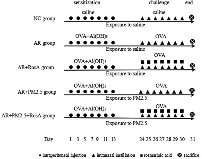
Experimental protocols applied in our study including induction of allergic rhinitis rat models, PM2.5 exposure, and rosmarinic acid intervention. The rats were randomly divided into 5 groups (8 animals per group). AR, allergic rhinitis model group; AR + PM2.5, allergic rhinitis with PM2.5 inhalation exposure model group; AR + PM2.5 + RosA, allergic rhinitis with RosA intervention for PM2.5 inhalation exposure model group; AR + RosA, allergic rhinitis with RosA intervention model group; NC, normal control group
2.5. Observation on nasal symptoms of AR rats
The frequency of sneezing and nasal rubs of all rats in various groups was recorded 15 min after the last nasal provocation on the 30th day. Filter strips of 1.0 × 25.0 mm2 were inserted into one of the nasal cavities of all rats to absorb nasal secretions for 5 min; then, the weight of nasal secretions (mg) was measured through weight‐reduction method. The above data were collected to evaluate nasal allergic symptoms of the rats. 22
2.6. Detection of expressions of IL‐4, IL‐13, INF‐γ in nasal lavage fluid, and OVA‐sIgE in serum through ELISA
After the assessment of nasal biological symptoms, rats in different groups were administered intraperitoneal injection of 3% pentobarbital sodium (30 mg/kg) to induce complete anesthesia. Then, nasal lavage fluid and blood samples from the abdominal aorta of the rat models were collected, centrifuged at 3,000 rpm for 10 min, and frozen at −80°C until further processing. ELISA kit (Xitang Biotechnology) was used to determine the expressions of IL‐4, IL‐13, and INF‐γ in the nasal lavage fluid, and that of OVA‐sIgE in serum, according to the manufacturer's instructions.
2.7. Observation on the pathological changes of nasal mucosa in rat models
The nasal mucosal tissue of all rat models was decalcified, embedded in paraffin, and cut into 4 mm sections. The sections were stained with hematoxylin–eosin (HE) stain to assess the pathological changes in the mucosa.
2.8. Detection of T‐bet and GATA‐3 mRNA expression in nasal mucosa through RT‐PCR
The extraction of total RNA from nasal mucosa of all rat models was conducted applying TRIzol reagent (Invitrogen) following the instructions from the manufacturer. The mRNA expressions of T‐bet and GATA‐3 in nasal mucosa were determined through SYBR green staining. The cDNA reverse transcription kit was applied for RT‐PCR detection and cDNA amplification. All primers were synthesized by Invitrogen, and their sequences are listed in Table 1. β‐actin was used as an endogenous reference. The relative mRNA levels of T‐bet and GATA‐3 were calculated by 2−ΔΔCt method.
TABLE 1.
Primer sequences of T‐bet and GATA‐3 applied for real‐time PCR
| Symbol | Primer (5′ to 3′) |
|---|---|
| β‐actin | Forward: 5′‐CCTCTATGCCAACACAGT‐3′ |
| Reverse: 5′‐ AGCCACCAATCCACACAG‐3′ | |
| T‐bet | Forward: 5′‐CCTGCTGGACGACAATGG‐3′ |
| Reverse: 5′‐ TCTGGTAGGCGGTGACTG‐3′ | |
| GATA−3 | Forward: 5′‐ AAGAGTGCCTCAAGTATCAG‐3′ |
| Reverse: 5′‐ GCGGATAGGTGGTAATGG‐3′ |
2.9. Detection of protein levels of NF‐κBp65 and IκBα in nasal mucosa through Western Blot
Total protein was extracted from the icy homogenate of the nasal mucosa from all rats. The protein levels of NF‐κB p65 and IκBα were determined through Western blot assay. The total protein samples were taken for electrophoresis, transferred to PVDF membrane, and incubated overnight with NF‐κB p65, IκBα, GAPDH, and histone H3 as the primary antibodies at 4°C. Subsequently, the membranes were incubated with labeled secondary antibodies from rabbit for another one hour. The protein bands were developed and fixed according to the instructions of the ECL kit. Then, the imaging of protein bands of all rat models was observed, and the gray value of the bands was analyzed by Quantity One software. Relative expression of the target protein equaled the gray value of the target protein/the gray value of internal reference protein.
2.10. Statistical analysis
The SPSS 20.0 software was applied for statistical analysis in the present study. All continuous variables are presented as mean ± standard deviation. Multi‐group comparisons were performed using one‐way analysis of variance (ANOVA) followed by Tukey post‐test. p values < 0.05 were considered indicative of statistical significance.
3. RESULTS
3.1. AR symptoms in rats
Rats in the AR group showed increased frequencies of sneezing and nasal itching, and the amount of nasal secretion was also significantly increased compared with those in the NC group (p < 0.001, Figure 2A–C). We also found that exposure to PM2.5 significantly worsened the above symptoms of AR rats when compared to those of the AR group (p < 0.001), while RosA intervention alleviated the symptoms of rats in the AR + RosA group (p < 0.001). Taking the two factors into consideration, we discovered that the above negative effects in rats in the AR + PM2.5 + RosA group were reversed after treatment with RosA for PM2.5 exposure (p < 0.001).
FIGURE 2.
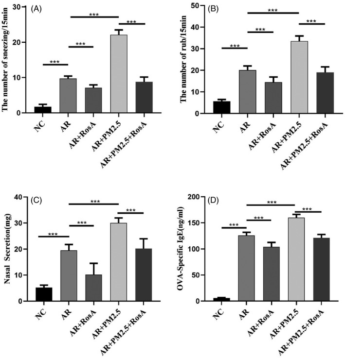
Effect of rosmarinic acid on nasal symptoms and serum levels of OVA‐specific IgE in PM2.5‐exposed allergic rhinitis rat models (N = 8 per group). AR, allergic rhinitis model group; AR + PM2.5 + RosA, allergic rhinitis with RosA intervention for PM2.5 inhalation exposure model group; AR + RosA, allergic rhinitis with RosA intervention model group; AR + PM2.5, allergic rhinitis with PM2.5 inhalation exposure model group; NC, normal control group. *p < 0.05, **p < 0.01, ***p < 0.001
3.2. Effects of RosA on mucosal histopathology in PM2.5‐exposed AR rats
Pathological examination of the nasal mucosa of rats in the AR group revealed disorganization of the epithelium, cell exfoliation, eosinophilic infiltration of lamina propria, gland swelling, and submucosal vascular congestion, when compared to those in the NC group (Figure 3A,B, the NC and AR groups). Similarly, PM2.5 exposure aggravated the above pathological manifestations (Figure 3D, the AR + PM2.5 group), while intervention with RosA led to reduced eosinophil infiltration, ordered epithelial arrangement, and reduced cell exfoliation in the nasal mucosa (Figure 3C, the AR + RosA group), and reversed the pathological manifestations caused by PM2.5 exposure (Figure 3E, the AR + PM2.5 + RosA group).
FIGURE 3.
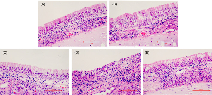
Effect of rosmarinic acid on the histopathological changes in nasal mucosa of PM2.5‐exposed allergic rhinitis rat models. Pathological examination of the nasal mucosa of rats in the (A) NC group; (B) AR group, (C) AR + RosA group, (D) AR + PM2.5 group, and (E) AR + PM2.5 + RosA group. AR, allergic rhinitis model group; AR + PM2.5 + RosA, allergic rhinitis with RosA intervention for PM2.5 inhalation exposure model group (400×, Scale bar, 50 μm); AR + RosA, allergic rhinitis with RosA intervention model group; AR + PM2.5, allergic rhinitis with PM2.5 inhalation exposure model group; NC, normal control group
3.2.1. Effects of RosA on OVA‐specific IgE levels in PM2.5‐exposed AR rats
We observed an obvious increase in the levels of serum OVA‐sIgE in the AR groups, when compared to those of the NC group (p < 0.001, Figure 2D). Moreover, in comparison with those of the AR group, the levels of OVA‐sIgE in the AR + PM2.5 rats were significantly upregulated (p < 0.001, Figure 2D). On the contrary, a significant decrease in the levels of serum OVA‐sIgE in the AR + RosA rats was observed when compared with those of the AR group (p < 0.001, Figure 2D). In addition, down‐regulated expressions of OVA‐sIgE were noted in rats of the AR + PM2.5 + RosA group after RosA intervention when compared to those in the AR + PM2.5 group (p < 0.001, Figure 2D).
3.2.2. Effects of RosA on IFN‐γ, IL‐4, and IL‐13 levels in PM2.5‐exposed allergic rhinitis rats
Compared with the NC group, rats in the AR group showed significantly decreased expression of IFN‐γ and increased expressions of IL‐4 and IL‐13 (p < 0.001, Figure 4). Moreover, the changes in the expressions of the above cytokines seemed to be aggravated by exposure to PM2.5 in the AR + PM2.5 group, in comparison with those in the AR group (p < 0.001, Figure 4). Nevertheless, RosA treatment in the AR + RosA group alleviated the expression trends of the above cytokines in comparison with the AR group (p < 0.01, Figure 4). In comparison with rats in the AR + PM2.5 group, rats in the AR + RosA + PM2.5 group showed a reverse trend of the above cytokines after RosA treatment for PM2.5 exposure (p < 0.001, Figure 4).
FIGURE 4.
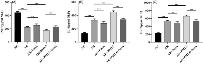
Effect of rosmarinic acid on the expression levels of IFN‐γ, IL‐4, and IL‐13 in nasal lavage fluid of PM2.5‐exposed allergic rhinitis rat models (N = 8 per group). AR, allergic rhinitis model group; AR + PM2.5, allergic rhinitis with PM2.5 inhalation exposure model group; AR + PM2.5 + RosA, allergic rhinitis with RosA intervention for PM2.5 inhalation exposure model group; AR + RosA, allergic rhinitis with RosA intervention model group; NC, normal control group. *p < 0.05, **p < 0.01, ***p < 0.001
3.2.3. Effects of RosA on the mRNA expression of T‐bet and GATA‐3 in PM2.5‐exposed allergic rhinitis rats
The mRNA expression of T‐bet in the nasal mucosa in the AR group decreased, while that of GATA‐3 increased markedly as compared to those in the NC group (p < 0.001, Figure 5). Moreover, PM2.5 exposure worsened the trend of changes in the expressions of T‐bet and GATA‐3 mRNA compared with those of rats in the AR group (p < 0.05, Figure 5, the AR + PM2.5 group), while RosA intervention alleviated the trend of changes (p < 0.01, Figure 5, the AR + RosA group). Moreover, RosA intervention reversed the damage caused by PM2.5 exposure regarding the expressions of the above genes in rats of the AR + PM2.5 + RosA group (p < 0.001, Figure 5, the AR + PM2.5 + RosA group).
FIGURE 5.

Effect of rosmarinic acid on the expression levels of T‐bet and GATA‐3 mRNA in the nasal mucosa of PM2.5‐exposed allergic rhinitis rat models (N = 8 per group). AR, allergic rhinitis model group; AR + PM2.5 + RosA, allergic rhinitis with RosA intervention for PM2.5 inhalation exposure model group; AR + RosA, allergic rhinitis with RosA intervention model group; AR + PM2.5, allergic rhinitis with PM2.5 inhalation exposure model group; NC, normal control group. *p < 0.05, **p < 0.01, ***p < 0.001
3.2.4. Effects of RosA on the NF‐κB signaling pathway in PM2.5‐exposed allergic rhinitis rats
Similarly, the protein expression of NF‐κB p65 in the nuclei of nasal mucosa cells of AR rats increased, while that of IκBα in cytoplasm decreased when compared to those of rats in the NC group (p < 0.001, Figure 6). PM2.5 exposure made the above changes more obvious in comparison with those of rats in the AR group (p < 0.001, Figure 6), whereas RosA intervention reversed the above trend of changes in the protein expression of NF‐κB p65 in cell nuclei and IκBα in cytoplasm, when compared to those of rats in the AR group and AR + PM2.5 group (both p < 0.01, Figure 6).
FIGURE 6.

Effect of rosmarinic acid on the expression levels of IκBα (in cytoplasm) and NF‐κB p65 (in cell nuclei) in nasal mucosa of PM2.5‐exposed allergic rhinitis rat models (N = 8 per group). AR + PM2.5, allergic rhinitis with PM2.5 inhalation exposure model group; AR + PM2.5 + RosA, allergic rhinitis with RosA intervention for PM2.5 inhalation exposure model group; AR + RosA, allergic rhinitis with RosA intervention model group; NC, normal control group; AR, allergic rhinitis model group. *p < 0.05, **p < 0.01, ***p < 0.001
4. DISCUSSION
Rosmarinic acid is considered as a promising alternative treatment for allergic diseases including AR, owing to its anti‐inflammatory, anti‐allergic, antibacterial, and antitumor properties. 23 , 24 , 25 Severe air pollution is associated with increased prevalence and severity of AR through numerous allergy‐related pollutants including PM2.5 particles. 3 , 6 , 7 In our study, we observed increased expression of OVA‐sIgE in AR rats, especially after PM2.5 exposure, which showed a positive correlation with the occurrence of allergic nasal symptoms. RosA was found to reduce the nasal irritation and down‐regulate the expression of OVA‐sIgE in the AR rat models. We also discovered that RosA intervention improved these symptoms in PM2.5‐exposed AR rats and significantly decreased the level of OVA‐sIgE in serum of AR rats; however, RosA intervention could not completely cure the allergy symptoms. These results indicate that RosA reduce the severity of nasal symptoms in AR rats exposed to PM2.5 and may alleviate allergic response by reducing the expression of allergy‐related factors such as OVA‐sIgE.
Imbalance of Th1/Th2 pattern is believed to play a key role in the pathophysiology of AR, which is characterized by an enhanced Th2 immune response. 26 , 27 Moreover, IL‐4 and IL‐13 are typical Th2 type cytokines which stimulate Th2 differentiation and IgE production by B cells. These have been reported to play a key role in allergic airway inflammation 28 through their participation in Th2 inflammatory response. 29 , 30 T‐bet and GATA‐3 are transcription factors of Th1 and Th2, respectively. Consistent with previous studies, we verified an imbalance of Th1/Th2 pattern in AR groups compared with the NC group. As an important air pollutant, PM2.5 enhances the antigenicity of allergens and aggravates allergic reactions in the body. 31 Moreover, in previous studies, PM2.5 was shown to significantly aggravate the nasal symptoms of AR patients and promote the development of nasal mucositis. 32 In the present study, we confirmed that PM2.5 exposures aggravated allergic symptoms and type 2 inflammation in AR rat models while it weakened type 1 inflammation.
As previously reported, the myriad effects of RosA and its derivatives are mediated via modulation of different signal transduction pathways and biological functions. 33 , 34 Specifically, its anti‐inflammatory and immunomodulatory effects have been confirmed in many studies. In the study by Jang et al, RosA was found to inhibit the symptoms of atopic dermatitis mice induced by 2,4‐dinitrofluorobenzene through suppressing the expression of IL‐4, and reducing the level of total IgE in serum. 35 Our present results showed that RosA could up‐regulate the expressions of Th1‐related cytokines and T‐bet mRNA, and down‐regulate Th2‐related cytokines and GATA‐3 mRNA. Therefore, we hypothesized that RosA may improve AR symptoms via regulating the balance of Th1/Th2 cytokines. In our study, RosA was found to reduce the expressions of type 2 cytokines (IL‐4 and IL‐13) and increase the expressions of type 1 cytokines (INF‐γ) when compared to those in the PM2.5 exposure groups. Moreover, the histopathological findings of PM2.5‐exposed AR rat model after RosA intervention revealed that RosA may induce rearrangement of the cilia and reduce the infiltration of the interstitial cells and the eosinophils. Our results indicated that RosA may inhibit the type 2 immune response and enhance type 1 immune response in PM2.5‐exposed AR rat models. These findings suggested a potential therapeutic role of RosA in PM2.5‐exposed AR; however, further studies are required to assess the clinical effectiveness and the underlying mechanism.
NF‐κB is an important intracellular nuclear transcription factor, which is involved in inflammatory reactions and immune responses. The main steps in the activation of the NF‐κB pathway include the phosphorylation, ubiquitination and degradation of IκBα, and the nuclear translocation of activated NF‐κB p65. 36 In our study, PM2.5‐exposed AR rats showed increased protein expression of NF‐κB p65 in the nuclei of nasal mucosal cells and decreased expression of IκBα in the cytoplasm. This indicates that PM2.5 exposure induced activation of the NF‐κB pathway. After RosA intervention, the nuclear shift of NF‐κB and the degradation of IκBα was reduced in PM2.5‐exposed AR rat models. RosA was shown to reduce lung injury in mice asthma models by suppressing the activation of the NF‐κB signaling pathway. 37 Similarly, RosA also reduced lung injury in mice asthma models by suppressing the activation of the NF‐κB signaling pathway. NF‐κB has been shown to play an important role in the production of Th2 cytokine in AR models. 38 Therefore, we speculate that RosA may alleviate the severity of PM2.5‐exposed AR through regulation of the NF‐κB signaling pathway and modulation of the balance of Th1/Th2 response.
However, there are several limitations in our study. There are a great variety of air pollutants with complex interactions among them. The compositions of PM2.5 may change with time and may differ in different districts. This posed a challenge in precisely replicating the real‐time atmospheric environment. Therefore, multi‐center experimental studies may be required to further explore and demonstrate the inflammatory reaction of PM2.5 exposure to AR models. Further clinical studies are also required to validate the effectiveness of RosA intervention in patients with AR after PM2.5 exposure.
Our findings suggest that RosA may improve allergic symptoms and reverse morphological changes in PM2.5‐exposed AR rat models. The possible mechanism of action of RosA may involve modulation of the expression of Th1/Th2 cytokines and the activation of the NF‐κB pathway. Our findings may provide new insights for the prevention of AR exacerbated by PM2.5 exposure.
CONFLICT OF INTEREST
We declare that we have no conflict of interest.
Zhou L, Huang Y, Han Z, et al. Effects of rosmarinic acid on the inflammatory response in allergic rhinitis rat models after PM2.5 exposure. J Clin Lab Anal. 2022;36:e24316. doi: 10.1002/jcla.24316
Funding information
This work was supported by the National Natural Science Foundation of China (No. 81670906, 81371078)
DATA AVAILABILITY STATEMENT
The datasets used and/or analyzed during the current study are available from the corresponding author on reasonable request.
REFERENCES
- 1. Cheng L, Chen J, Fu Q, et al. Chinese society of allergy guidelines for diagnosis and treatment of allergic rhinitis. Allergy Asthma Immunol Res. 2018;10:300‐353. [DOI] [PMC free article] [PubMed] [Google Scholar]
- 2. Meltzer EO. Allergic rhinitis: burden of illness, quality of life, comorbidities, and control. Immunol Allergy Clin North Am. 2016;36:235‐248. [DOI] [PubMed] [Google Scholar]
- 3. Lee J, Yun S, Oh I. Impact of environmental factors on the prevalence changes of allergic diseases in elementary school students in Ulsan, Korea: a longitudinal study. Int J Environ Res Public Health. 2020;17(23):8831. [DOI] [PMC free article] [PubMed] [Google Scholar]
- 4. Tamay Z, Akcay A, Ones U, Guler N, Kilic G, Zencir M. Prevalence and risk factors for allergic rhinitis in primary school children. Int J Pediatr Otorhinolaryngol. 2007;71:463‐471. [DOI] [PubMed] [Google Scholar]
- 5. Todkill D, de Jesus Colon Gonzalez F, Morbey R,, et al. Environmental factors associated with general practitioner consultations for allergic rhinitis in London, England: a retrospective time series analysis. BMJ Open. 2020;10(12):e036724. [DOI] [PMC free article] [PubMed] [Google Scholar]
- 6. Li CH, Sayeau K, Ellis AK. Air pollution and allergic rhinitis: role in symptom exacerbation and strategies for management. J Asthma Allergy. 2020;13:285‐292. [DOI] [PMC free article] [PubMed] [Google Scholar]
- 7. Pinkerton KE, Green FH, Saiki C, et al. Distribution of particulate matter and tissue remodeling in the human lung. Environ Health Perspect. 2000;108:1063‐1069. [DOI] [PMC free article] [PubMed] [Google Scholar]
- 8. Woo KS, Chook P, Hu YJ, et al. The impact of particulate matter air pollution (PM2.5) on atherosclerosis in modernizing China: a report from the CATHAY study. Int J Epidemiol. 2021;50:578‐588. [DOI] [PubMed] [Google Scholar]
- 9. García‐Serna AM, Hernández‐Caselles T, Jiménez‐Guerrero P, et al. Air pollution from traffic during pregnancy impairs newborn's cord blood immune cells: the NELA cohort. Environ Res. 2021;198:110468. [DOI] [PubMed] [Google Scholar]
- 10. Wang M, Wang S, Wang X, et al. The association between PM(2.5) exposure and daily outpatient visits for allergic rhinitis: evidence from a seriously air‐polluted environment. Int J Biometeorol. 2020;64:139‐144. [DOI] [PubMed] [Google Scholar]
- 11. Guo ZQ, Dong WY, Xu J, et al. T‐helper type 1‐T‐helper type 2 shift and nasal remodeling after fine particulate matter exposure in a rat model of allergic rhinitis. Am J Rhinol Allergy. 2017;31:148‐155. [DOI] [PubMed] [Google Scholar]
- 12. Kim GD, Park YS, Jin YH, Park CS. Production and applications of rosmarinic acid and structurally related compounds. Appl Microbiol Biotechnol. 2015;99:2083‐2092. [DOI] [PubMed] [Google Scholar]
- 13. Hasanein P, Seifi R. Beneficial effects of rosmarinic acid against alcohol‐induced hepatotoxicity in rats. Can J Physiol Pharmacol. 2018;96:32‐37. [DOI] [PubMed] [Google Scholar]
- 14. Değer U, Çavuş Y. Investigation of the role of rosmarinic acid treatment in regulating inflammation, cell damage, and angiogenesis in rat ovarian torsion and detorsion models. Acta Cir Bras. 2020;35:e202000304. [DOI] [PMC free article] [PubMed] [Google Scholar]
- 15. Alagawany M, Abd El‐Hack ME, Farag MR, et al. Rosmarinic acid: modes of action, medicinal values and health benefits. Anim Health Res Rev. 2017;18:167‐176. [DOI] [PubMed] [Google Scholar]
- 16. Messeha SS, Zarmouh NO, Asiri A, Soliman KFA. Rosmarinic acid‐induced apoptosis and cell cycle arrest in triple‐negative breast cancer cells. Eur J Pharmacol. 2020;885:173419. [DOI] [PMC free article] [PubMed] [Google Scholar]
- 17. Lv R, Du L, Liu X, Zhou F, Zhang Z, Zhang L. Rosmarinic acid attenuates inflammatory responses through inhibiting HMGB1/TLR4/NF‐κB signaling pathway in a mouse model of Parkinson's disease. Life Sci. 2019;223:158‐165. [DOI] [PubMed] [Google Scholar]
- 18. Khalaf AA, Hassanen EI, Ibrahim MA. Rosmarinic acid attenuates chromium‐induced hepatic and renal oxidative damage and DNA damage in rats. J Biochem Mol Toxicol. 2020;34:e22579. [DOI] [PubMed] [Google Scholar]
- 19. Hong Z, Guo Z, Zhang R, et al. Airborne fine particulate matter induces oxidative stress and inflammation in human nasal epithelial cells. Tohoku J Exp Med. 2016;239:117‐125. [DOI] [PubMed] [Google Scholar]
- 20. Guo Z, Hong Z, Dong W, et al. PM(2.5)‐induced oxidative stress and mitochondrial damage in the nasal mucosa of rats. Int J Environ Res Public Health. 2017;14(2):134. [DOI] [PMC free article] [PubMed] [Google Scholar]
- 21. Chu X, Ci X, He J, et al. Effects of a natural prolyl oligopeptidase inhibitor, rosmarinic acid, on lipopolysaccharide‐induced acute lung injury in mice. Molecules. 2012;17:3586‐3598. [DOI] [PMC free article] [PubMed] [Google Scholar]
- 22. Lindell E, Svensjö ME, Malm L, Petersson G. Tachykinin‐induced nasal fluid secretion and plasma exudation in the rat: effects of peptidase inhibition. Neuropeptides. 1995;28:309‐315. [DOI] [PubMed] [Google Scholar]
- 23. Babaei M, Borja Zamfir GM, Chen X, et al. Metabolic engineering of saccharomyces cerevisiae for rosmarinic acid production. ACS Synth Biol. 2020;9:1978‐1988. [DOI] [PMC free article] [PubMed] [Google Scholar]
- 24. Zhu F, Asada T, Sato A, Koi Y, Nishiwaki H, Tamura H. Rosmarinic acid extract for antioxidant, antiallergic, and α‐glucosidase inhibitory activities, isolated by supramolecular technique and solvent extraction from Perilla leaves. J Agric Food Chem. 2014;62:885‐892. [DOI] [PubMed] [Google Scholar]
- 25. Oh HA, Park CS, Ahn HJ, Park YS, Kim HM. Effect of Perilla frutescens var. acuta Kudo and rosmarinic acid on allergic inflammatory reactions. Exp Biol Med (Maywood). 2011;236:99‐106. [DOI] [PubMed] [Google Scholar]
- 26. Wang M, Zhang W, Shang J, Yang J, Zhang L, Bachert C. Immunomodulatory effects of IL‐23 and IL‐17 in a mouse model of allergic rhinitis. Clin Exp Allergy. 2013;43:956‐966. [DOI] [PubMed] [Google Scholar]
- 27. Eifan AO, Furukido K, Dumitru A, et al. Reduced T‐bet in addition to enhanced STAT6 and GATA3 expressing T cells contribute to human allergen‐induced late responses. Clin Exp Allergy. 2012;42:891‐900. [DOI] [PubMed] [Google Scholar]
- 28. Shirkani A, Mansouri A, Farid Hosseini R, et al. The role of interleukin‐4 and 13 gene polymorphisms in allergic rhinitis: a case control study. Rep Biochem Mol Biol. 2019;8:111‐118. [PMC free article] [PubMed] [Google Scholar]
- 29. Moh'd Al‐Rawashdeh B, Sada Alhanjori A, Ali E, Zihlif M. Association of IL‐4 polymorphisms with allergic rhinitis in jordanian population. Medicina. 2020;56(4):179. [DOI] [PMC free article] [PubMed] [Google Scholar]
- 30. Miyahara S, Miyahara N, Matsubara S, Takeda K, Koya T, Gelfand EW. IL‐13 is essential to the late‐phase response in allergic rhinitis. J Allergy Clin Immunol. 2006;118:1110‐1116. [DOI] [PubMed] [Google Scholar]
- 31. Minai‐Fleminger Y, Levi‐Schaffer F. Mast cells and eosinophils: the two key effector cells in allergic inflammation. Inflamm Res. 2009;58:631‐638. [DOI] [PubMed] [Google Scholar]
- 32. Steerenberg PA, Withagen CE, van Dalen WJ, et al. Adjuvant activity of ambient particulate matter of different sites, sizes, and seasons in a respiratory allergy mouse model. Toxicol Appl Pharmacol. 2004;200:186‐200. [DOI] [PubMed] [Google Scholar]
- 33. Naclerio R, Ansotegui IJ, Bousquet J, et al. International expert consensus on the management of Allergic Rhinitis (AR) aggravated by air pollutants: impact of air pollution on patients with AR: current knowledge and future strategies. World Allergy Organ J. 2020;13:100106. [DOI] [PMC free article] [PubMed] [Google Scholar]
- 34. Lin LZ, Chen HH. Rosmarinic acid protects on rat bone marrow mesenchymal stem cells from hydrogen peroxide‐induced apoptosis. J Asian Nat Prod Res. 2018;20:570‐580. [DOI] [PubMed] [Google Scholar]
- 35. Rong H, Liang Y, Niu Y. Rosmarinic acid attenuates β‐amyloid‐induced oxidative stress via Akt/GSK‐3β/Fyn‐mediated Nrf2 activation in PC12 cells. Free Radic Biol Med. 2018;120:114‐123. [DOI] [PubMed] [Google Scholar]
- 36. Williams LM, Gilmore TD. Looking down on NF‐κB. Mol Cell Biol. 2020;40:e00104‐e00120. [DOI] [PMC free article] [PubMed] [Google Scholar]
- 37. Liang Z, Nie H, Xu Y, et al. Therapeutic effects of rosmarinic acid on airway responses in a murine model of asthma. Int Immunopharmacol. 2016;41:90‐97. [DOI] [PubMed] [Google Scholar]
- 38. Fan XL, Zeng QX, Li X, et al. Induced pluripotent stem cell‐derived mesenchymal stem cells activate quiescent T cells and elevate regulatory T cell response via NF‐κB in allergic rhinitis patients. Stem Cell Res Ther. 2018;9:170. [DOI] [PMC free article] [PubMed] [Google Scholar]
Associated Data
This section collects any data citations, data availability statements, or supplementary materials included in this article.
Data Availability Statement
The datasets used and/or analyzed during the current study are available from the corresponding author on reasonable request.