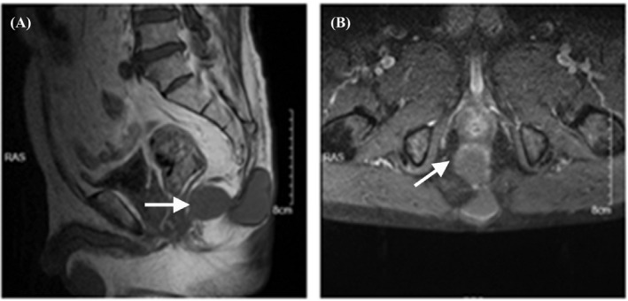
FIGURE 1.

High‐resolution computed tomography (HRCT) of the sacrococcygeal vertebrae showed oval cystic lesions involving in subcutaneous tissue and pelvic cavity. Figure A and Figure B represented the location and size of the abscess under the lateral position and frontal position, respectively. There was an oval cystic lesion near the fifth cone of sacrum and subcutaneous tissue, respectively, and the two lesions were connected. The size of the larger abscess was about 50 × 30 mm, and the size of the smaller abscess was about 40 × 30 mm. White arrow indicated the abscesses
