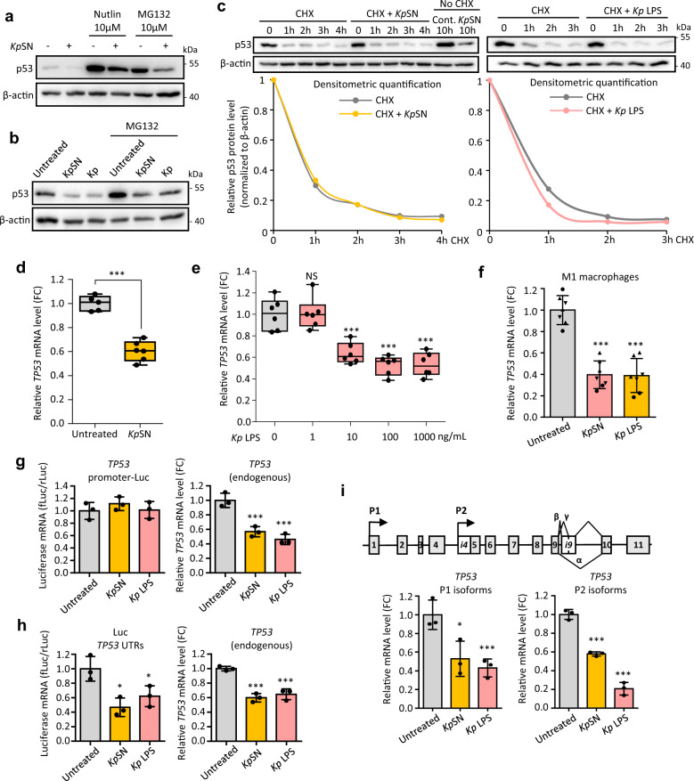
Fig. 5. p53 inhibition occurs at mRNA level.
a Downregulation of p53 protein level induced by KpSN in BJ hTert cells is not rescued by MDM2 inhibitor Nutlin or by proteasome inhibitor MG132. b Proteasome inhibition by MG132 does not rescue p53 downregulation upon direct infection of BJ hTert cells by K. pneumoniae (m.o.i. 10). c p53 protein stability in BJ hTert cells exposed to KpSN (orange) or 100 ng/mL LPS (pink) was investigated using cycloheximide (CHX) chase assay. Cells were pretreated 4 h with KpSN before CHX treatment. Lower panels show the densitometry quantification of Western blots bands normalized to β-actin level. d RT-qPCR for TP53 mRNA level of BJ hTert cells upon 8 h exposure to KpSN, assessed by. e, RT-qPCR for TP53 mRNA level of BJ hTert cells upon 8 h exposure to indicated dose of LPS. f RT-qPCR for TP53 mRNA level upon KpSN or 100 ng/mL LPS (8 h) in PBMC-derived macrophages, polarized into M1 phenotype. The different data point symbols indicate different blood donors. g Luciferase reporter assay using TP53 promoter-Luc construct transfected in BJ hTert cells. Transfection rate was normalized by co-transfection of Renilla luciferase vector. Endogenous TP53 mRNA level was measured in the same samples. h Luciferase reporter assay using a construct expressing the luciferase flanked by p53 UTRs, transfected in BJ hTert cells. Transfection rate was normalized by co-transfection of Renilla luciferase vector. Endogenous TP53 mRNA level was measured in the same samples. i Upper panel: structure of the TP53 gene with the two alternative promoters P1 and P2. Lower panel: mRNA level of P1 and P2 p53 isoforms in BJ hTert cells upon treatment with KpSN or 100 ng/mL LPS (8 h). *p < 0.05; ***p < 0.01; NS non-significant.

