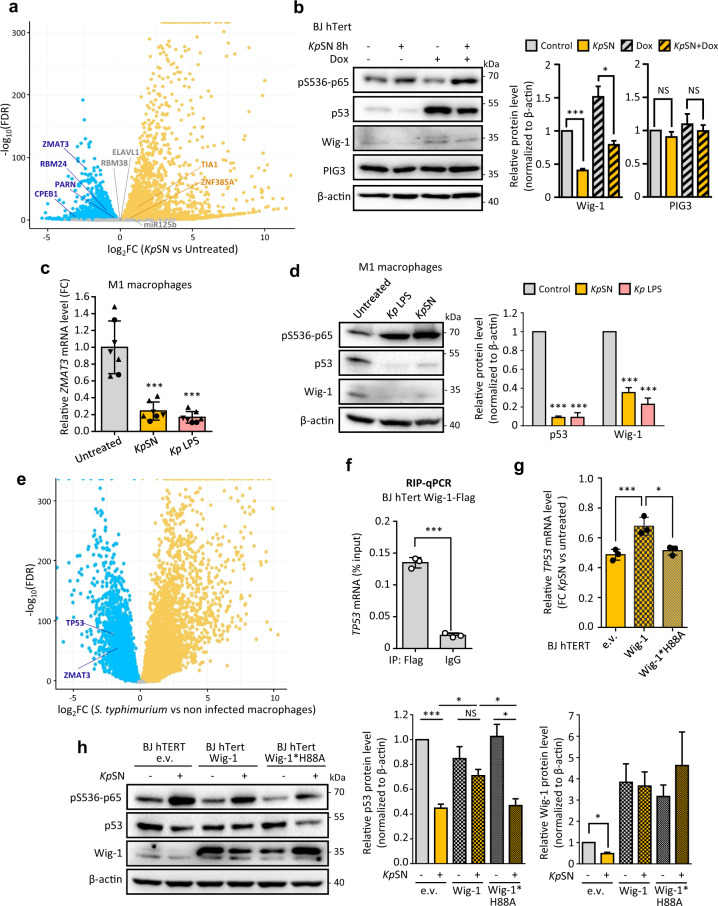
Fig. 6. LPS-induced downregulation of ZMAT3 destabilizes TP53 mRNA.
a Differential expression level of known factors regulating TP53 3ʹUTR in our RNA-seq data of KpSN-treated BJ hTert cells. b Left panel: Western blot of BJ hTert cells upon 8 h treatment with KpSN and doxorubicin. Right panel: densitometric quantification of Wig-1 and PIG3 Western blot bands normalized to β-actin level. c RT-qPCR for ZMAT3 mRNA level upon KpSN or 100 ng/mL LPS (8 h) in PBMC-derived macrophages, polarized into M1 phenotype. The different data point symbols indicate different blood donors. d Left panel: Western blot of M1 macrophages upon KpSN or LPS treatment. Right panel: densitometric quantification of p53 and Wig-1 Western blot bands normalized to β-actin level. e mRNA level of TP53 and ZMAT3 in RNA-seq data from PBMC-derived macrophages from 60 different blood donors infected by Salmonella thyphimurium. f Validation of Wig-1 binding to TP53 mRNA in BJ hTert cells by RIP-qPCR. g Rescue of KpSN-induced p53 downregulation in BJ hTert cells by Wig-1 overexpression but not by the RNA-binding deficient H88A mutant, assessed by RT-qPCR. h Left panel: Western blot of Wig-1 and Wig-1*H88A overexpressing BJ hTert cells upon KpSN treatment. Right panels: densitometric quantification of p53 and Wig-1 Western blot bands normalized to β-actin level. *p < 0.05; ***p < 0.01; NS non-significant.

