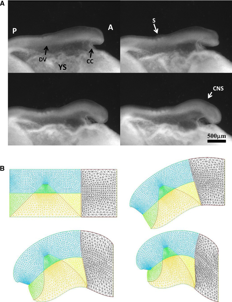
Fig. 10.
a In vivo time-lapse imaging of chicken flexure during the ventral pull, showing how the head part flexes ventrally (from Video 15). The movie shows the contraction of the cardiac crescent, and the forward flexure of the head. boundary; YSYolk Sac; AAnterior; PPosterior; CNSCentral Nervous System; SSomites; CCCardiac Crescent b Example of pattern built with a “mouth generating” contraction, an “ear generating” contraction and a “ventral” contraction (Video 16)

