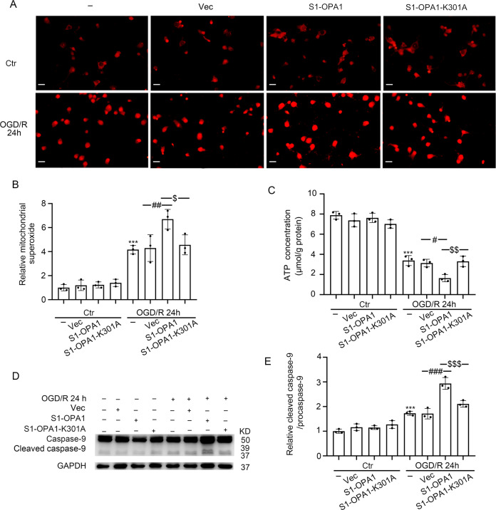
Fig. 4. S1-OPA1 overexpression induced mitochondrial superoxide generation, mitochondrial bioenergetic deficits, and mitochondrial apoptosis after OGD/R.
A Representative images of Mito-Sox staining in different overexpression groups under normal condition and OGD/R for 24 h. B Quantitative analysis of superoxide generation in neurons through red fluorescence intensity. C ATP content in neurons of each group was leveled by protein concentration. D Western blot analysis of caspase-9 and cleaved caspase-9 in neurons. E Ratio of cleaved caspase-9 to caspase-9 was quantitatively analyzed to represent the level of mitochondrial apoptosis. ***p < 0.001 vs. control group, ###p < 0.001, ##p < 0.01, #p < 0.05, $$$p < 0.001, $$p < 0.01, $p < 0.05; scale bar = 20 μm; n = 3.

