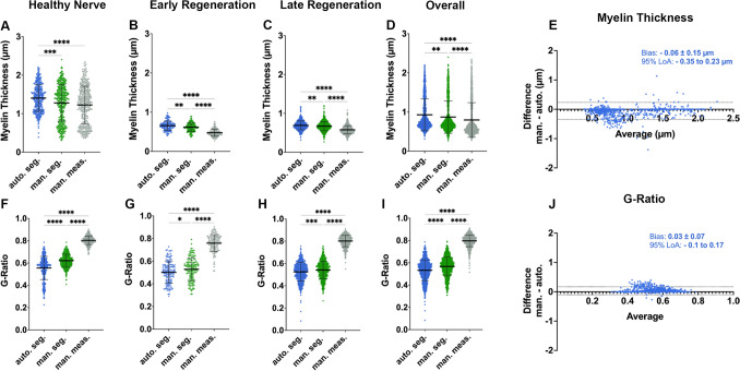
Figure 4.
Automated myelin sheath thickness measurements and g-ratio calculations. (A) Comparison of myelin sheath thickness results obtained from automated segmentations (blue), ground truth labels (green) and manual morphometry (grey) for healthy nerves, (B) early nerve regeneration stages, (C) late regeneration stages and (D) all samples combined. (E) Nerve fiber specific comparison in a bland–Altman plot showing good agreement of the AxonDeepSeg automated histomorphometry (auto.), with myelin thickness measurements in the ground truth (man.). We observed a tendency of AxonDeepSeg to overestimate the myelin thickness by 0.06 ± 0.15 µm on average (bias) with the 95% limits of agreement (LoA) being -0.1 µm to 0.17 µm. (F) Comparison of the g-ratio calculations obtained from automated segmentations (blue), ground truth labels (green) and manual morphometry (grey) for healthy nerves, (G) early nerve regeneration stages, (H) late regeneration stages and (I) all samples combined. As a result of the axon diameter overestimation and underestimated myelin sheath thickness in manual measurements (grey), the g-ratios calculated from these metrics deviated drastically from the ground truth. (J) Nerve fiber specific comparison of the g-ratios in a bland–Altman plot showing an acceptable agreement of the automated histomorphometry via AxonDeepSeg (auto.) with the ground truth measurements (man).

