Figure 2.
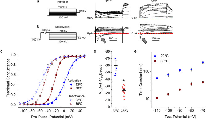
Physiological temperature reduces hERG 1a hysteresis of ionic currents. (a) Pulse protocol and corresponding sample ionic traces showing hERG 1a activation when stably expressed in HEK293 cells recorded at room temperature (22 °C, middle) or physiological temperature (36 °C, right). (b) Pulse protocol and corresponding sample ionic traces showing hERG 1a deactivation when stably expressed in HEK293 cells recorded at room temperature (22 °C, middle) or physiological temperature (36 °C, right). (c) Temperature-dependent shift in hERG 1a voltage dependence. Normalized peak tail current, representing fractional conductance, is plotted as a function of pre-pulse potential and fitted with a Boltzmann function for recordings completed at 22 °C (blue diamonds) and 36 °C (red squares). Channel activation recorded as in “(a)” is shown with solid symbols. Channel deactivation recorded as in “(b)” is shown with open symbols. (d) The magnitude of hysteresis was quantified by subtracting the V1/2 of deactivation from the V1/2 of activation. Hysteresis was significantly smaller in recordings completed at 36 °C (n = 12) compared with recordings at 22 °C (n = 5). (e) Time constants representing the fast deactivation time course for hERG 1a current recordings completed at either 22 °C (blue) or 36 °C (red). We fit current decay (arrows in “b”) with a double exponential equation (Eq. 4) and reported the fast time constant (τ) as a function of test potential. All data are reported as mean ± SEM. * indicates p < 0.05.

