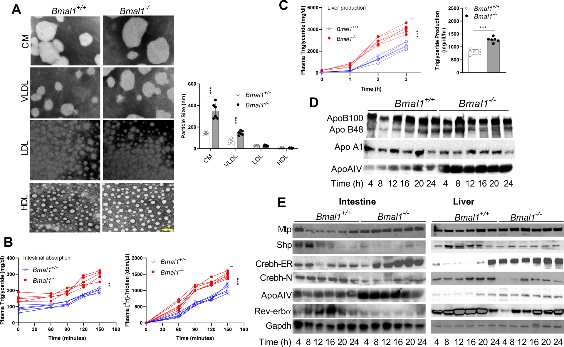
Figure 1: Hypertriglyceridemia and larger lipoproteins in chow-fed Bmal1−/− mice.

Bmal1−/− and Bmal1+/+ male siblings (12-week-old, ad libitum chow-fed) were sacrificed at 4 h intervals.
(A) Plasma collected at 12:00 was subjected to sequential density gradient ultracentrifugation. Isolated lipoproteins were negatively stained for electron microscopy (left). Scale bar = 50 nm. Diameters of different particles were quantified (right). Values are mean ± SD. ** p<0.01 and *** p<0.001, t-test.
(B) Mice fasted overnight were injected intraperitoneally with P407. After 1 h, they were gavaged with 3H-triolein in olive oil (50 μL) at 12:00. Blood was collected at the indicated times to measure triglyceride (left) and radioactivity (right). Two-way ANOVA, ** p<0.05, *** p<0.001.
(C) Mice fasted for 5 h were injected with P407 at 12:00, and plasma triglycerides were determined at different times. Time-dependent increases in plasma triglycerides (left) and triglyceride production rates (right) were significantly higher in Bmal1−/− than in Bmal1+/+ mice. Mean ± SD, n = 5–6 for each time point. Two-way ANOVA and t-test *** p<0.001.
(D) Plasma (1 μL) from different times was subjected to Western blotting to measure different apolipoproteins.
(E) Equal amounts of liver protein (25 μg) were used to detect proteins.
