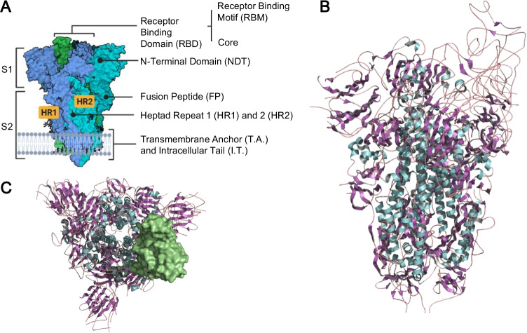
Fig. 1.
A Side view of the demarcating protein S2 and S1 domains and subdomains. B SARS-CoV-2 spike protein structure of the D614G mutant viral strain (PDB: 6XM0) constructed through PyMOL® and BioRender. The colors refer to the secondary structures: alpha-helix in cyan, beta sheets in purple and turns in magenta. C Top view of the trimeric protein with the receptor binding domain (RBD) demarcated on the surface

