
Figure 6.
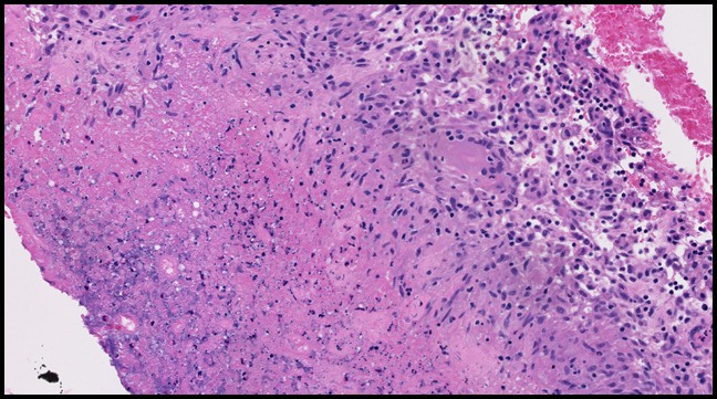
Photograph showing histologic examination of the preoperative ultrasonography-guided synovial biopsy demonstrated necrotic tissue that was bordered by lymphohistiocytic inflammation. This was not diagnostic for pigmented villonodular synovitis.