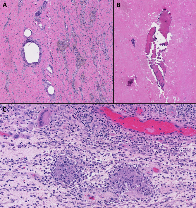
Figure 8.
Photographs showing histologic examination of the intraoperative periprosthetic tissue samples demonstrating detritic synovitis (A), abundant necrotic tissue with osteolysis (B), and noncaseating granulomatous inflammation (C). The samples were negative for Kinyoun and Fite stains. There were no features suggestive of pigmented villonodular synovitis.

