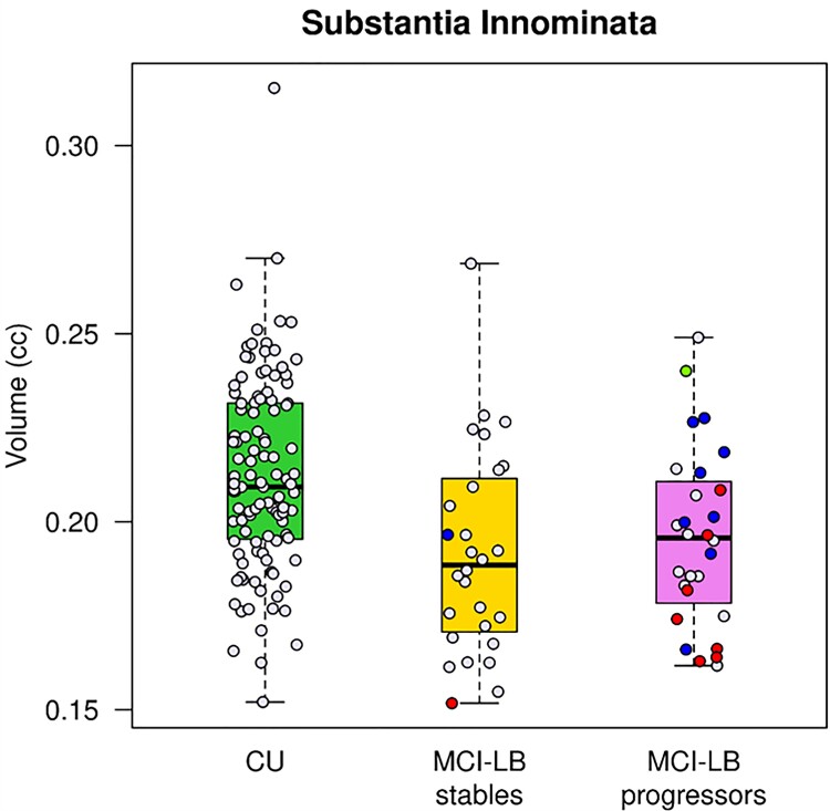
Figure 2.
Substantia innominata volumes. Box plots show that MCI-LB stables (FDR corrected P = 0.007) and MCI-LB progressors (FDR corrected P = 0.028) had smaller substantia innominata volumes compared to CU. The pathologically confirmed cases were colour coded based on the pathologic diagnosis after a median (range) of 5.3 (2.4–10.6) years. Blue labels represent cases with intermediate or high likelihood DLB (according to the fourth report of the DLB Consortium Criteria) who had low Alzheimer’s disease pathology (according to the NIA-AA criteria) classified as Lewy body disease. Red labels represent cases with intermediate or high likelihood DLB and intermediate or high Alzheimer’s disease, classified as having mixed Lewy body disease–Alzheimer’s disease pathology. The single green label represents a case with high Alzheimer’s disease and no Lewy body disease pathology classified as Alzheimer’s disease.

