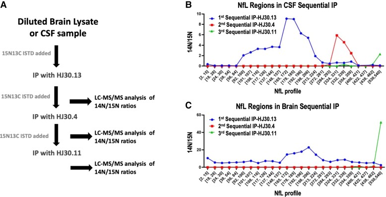
Figure 2.
Brain contains two main NfL species, whereas CSF has at least three main NfL species. Experimental method for sequential IP-MS/MS assay purifying and identifying at least three NfL fragment species (A). Sequential NfL IP from pooled CSF (n = 1) indicates three main NfL domains: a mid-domain region from NfL93 to NfL224, another region from NfL324 to NfL359 and a C-terminal region at NfL530 (B) and brain cortex lysate (n = 1) showing full-length NfL from NfL2 to NfL540, with a C-terminal peptide at NfL 530 (C). The blue line depicts peptides identified following the first IP with HJ30.13, the red line depicts peptides identified during the second IP with HJ30.4 and the green line represents peptides identified during the third IP with HJ30.11.

