Fig. 1.
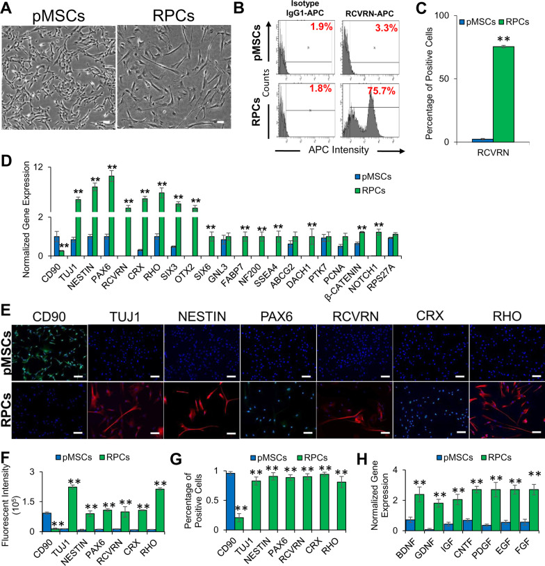
Characterization of RPCs derived from pMSCs. A Phase-contrast images of pMSCs and RPCs. 100 µm scale bar. (Magnification: ×4). B, C Histograms and graphical representation of the expression of the retinal marker, RCVRN, by pMSCs and RPCs as determined by flow cytometry (**p ≤ 0.01). D Expression of MSC (CD90), neural (TUJ1, NESTIN, and PAX6), retinal (RCVRN, CRX, RHO, SIX3, OTX2, SIX6, GNL3, FABP7, NF200, SSEA4, ABCG2, DACH1, PTK7, PCNA, β-CATENIN, NOTCH1, and RPS27A) genes in pMSCs and RPCs as determined by qRT-PCR. Gene expression was normalized to GAPDH and β-ACTIN. Error bars represent the SEM (**p ≤ 0.01). E Expression of MSC (CD90), neural (TUJ1, NESTIN, and PAX6), and retinal (RCVRN, CRX, and RHO) proteins in pMSCs and RPCs as visualized by immunocytostaining. Shown are representative merged images of DAPI (blue) and human antibodies (green and red). 100 µm scale bar (Magnification: ×10). F Measurement of fluorescent intensity of proteins immunocytostained using antibodies in pMSCs and RPCs (**p ≤ 0.01). G Percentage of positive immunocytostained cells (**p ≤ 0.01). H Expression of neurotrophic genes, BDNF, GDNF, IGF, CNTF, PDGF, EGF, and FGF in pMSCs and RPCs. Gene expression was normalized to GAPDH and β-ACTIN, and error bars represent the SEM (**p ≤ 0.01). Significant changes in the gene and protein expression were observed upon differentiation of pMSCs into RPCs. All experiments were carried out in triplicates

