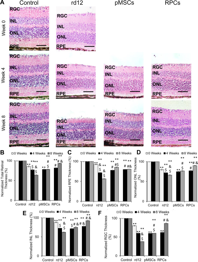
Fig. 5.
Histological analysis of rd12 retina transplanted with human cells. A H&E staining of paraffin-embedded sections of the control (wild-type), rd12, pMSCs and RPCs transplanted retina harvested at 0 week, 4 week, and 8 weeks. All scale bars represent 50 μm. (Magnification: ×40). B–F Graphical representation comparing the average thickness of the retina, RPE, ONL, INL, and RGC. Thickness of each retina was normalized to the control (wild-type) retina. Symbols, **, #, & and $ indicate significant difference at p ≤ 0.01 between all experimental conditions: control (wild-type), rd12, rd12 + pMSCs and rd12 + RPCs, respectively

