
Figure 3. Xenografted human cortical progenitors generate both excitatory and inhibitory cortical neurons in the same clone.
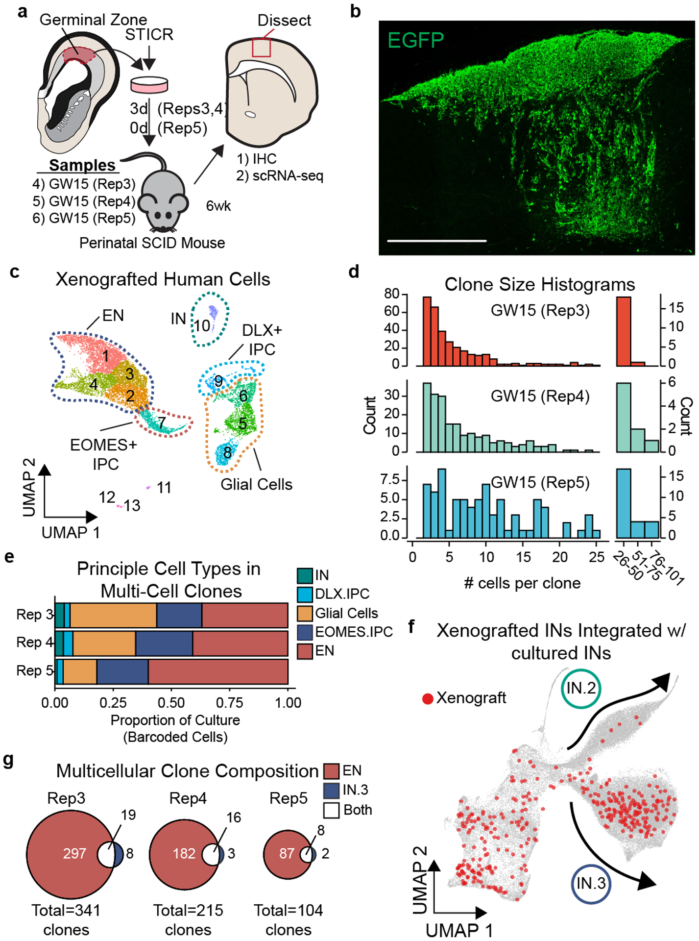
a) Schematic depicting experimental design and analysis of STICR-labeled progenitors by IHC and scRNA-seq following transplantation into the postnatal murine cortex. b) Representative image of transplanted human cortical cells. EGFP expression from STICR depicted in green. Scale bar, 500um. c) UMAP embedding and Louvain clustering of xenografted cells following scRNA-seq. d) Histogram of clone sizes within each xenograft sample. Left, clone sizes from 1–55 cells. Right, clone sizes of >25 cells in 25 cell bins. e) Stacked barplot depicting relative proportion of principal cell types within multi-cellular clones of each sample. f) UMAP embedding of both cultured and xenograft-derived INs. Xenograft-derived cells that are members of multicellular clones highlighted in red. IN.2 and IN.3 trajectories depicted with arrows. g) Venn diagram showing number of multi-cell clones containing excitatory neurons and/or IN.3 neurons.