
Figure 5.
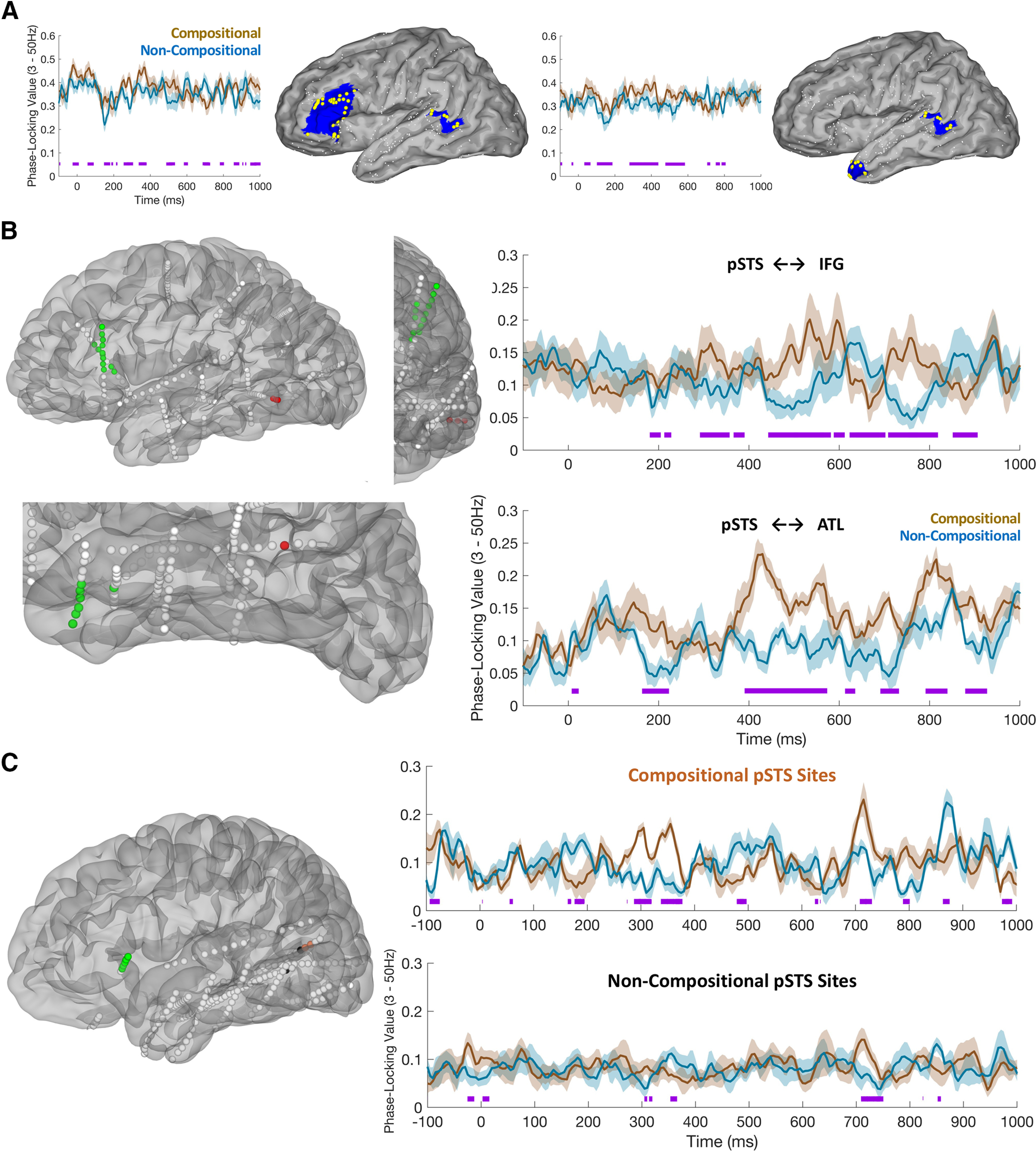
Phase locking between semantic composition regions of interest. A, Left, Average generalized phase-locking values (gPLV) for five patients showing greater gPLV for phrase composition relative to noncomposition between pSTS (HCP index, TPOJ1) and pars triangularis (HCP index, 45, IFSa, IFSp, 47l). Right, Average gPLVs for the six patients showing greater phase locking between pSTS and temporal pole (HCP index, TGd). Purple lines indicate points of significant conditional differences in gPLV values (FDR corrected for multiple comparisons). gPLV values are plotted from prestimulus baseline (−500 to −100 ms before first word onset). In the brain plots, small white spheres represent electrodes from across all patients that fall outside the region of interest; large yellow spheres represent electrodes included within the region of interest; blue shading represents the HCP surface corresponding to each region of interest. B, Posterior temporal lobe (pSTS) gPLVs with inferior frontal gyrus (specifically, pars triangularis, top) and anterior temporal lobe (specifically, temporal pole, bottom). Left, Plots show the localization in native space of electrodes significantly involved (q < 0.05) in interregional phase locking (3–50 Hz). Right, Plots show average time courses (mean ± SEM) of phase-locking value changes from baseline in phrase composition (brown) and noncomposition (blue) trials. C, Right, Phase-locking values between pSTS and pars triangularis in an exemplar patient, contrasting PLVs for electrodes in pSTS that showed an effect of phrase composition (top, orange electrodes) with those in pSTS that did not show an effect of composition (bottom, black electrodes). Because pSTS is a small ROI, only a subset of our patients satisfied the criteria for this analysis, that is, those (1) exhibiting joint coverage across pSTS and pars triangularis and (2) having electrodes in pSTS that did show an effect of composition and other electrodes that did not.