
Figure 6.
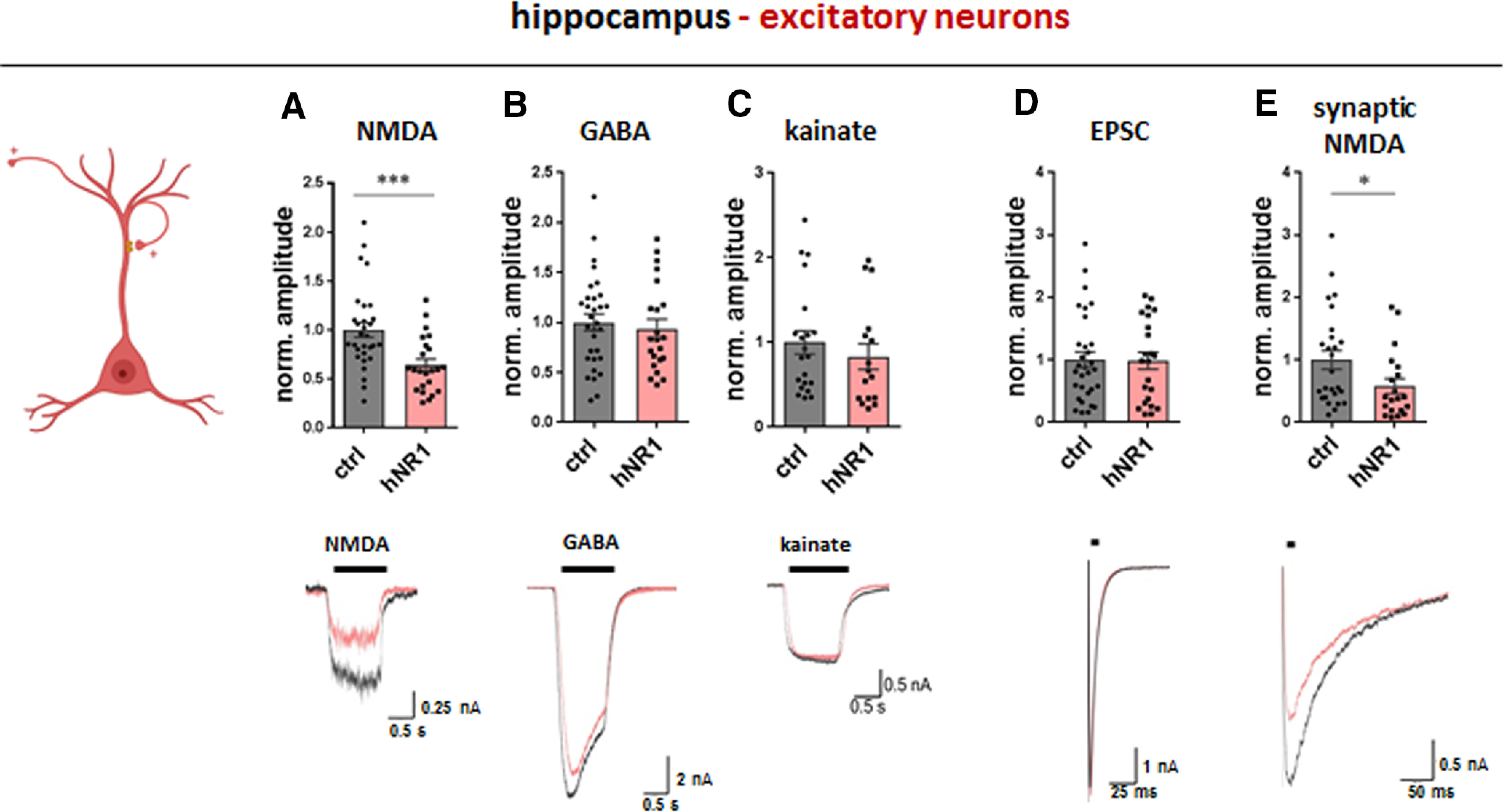
hNR1 antibody impairs NMDA currents of excitatory hippocampal neurons. In hippocampal autaptic cultures, 24-h treatment with hNR1 antibody (1 µg/ml) selectively decreases whole-cell (A) and synaptic (E) NMDA currents of excitatory neurons, when compared to treatment with control (ctrl) antibody. A–C, Whole-cell receptor currents were evoked by 1s bath application of (A) NMDA (10 μm), (B) GABA (30 μm), or (C) kainate (20 μm). Quantification of normalized current amplitudes from three independent experiments: (A) NMDA currents: ctrl = 1 ± 0.08, n = 30 neurons, hNR1 = 0.64 ± 0.06, n = 23 neurons, p = 0.0008; (B) GABA currents: ctrl = 1 ± 0.08, n = 31 neurons, hNR1 = 0.94 ± 0.09, n = 22 neurons; (C) kainate currents: ctrl = 1 ± 0.14, n = 21 neurons, hNR1 = 0.83 ± 0.15, n = 16 neurons. D, E, Synaptic responses were evoked by a brief somatic depolarization of neurons from −70 to 0 mV for 2 ms, synaptic NMDA currents were recorded in the presence of 0 mm Mg2+, 10 μm glycine, and 10 μm NBQX. Quantification of normalized current amplitudes from three independent experiments: (D) EPSCs: ctrl = 1 ± 0.13, n = 31 neurons, hNR1 = 0.99 ± 0.14, n = 23 neurons; (E) synaptic NMDA: ctrl = 1 ± 0.15, n = 26 neurons, hNR1 = 0.58 ± 0.12, n = 20 neurons, p = 0.039. Error bars indicate SEM. Unpaired t test was used to evaluate statistical significance. *p < 0.05, ***p < 0.001.