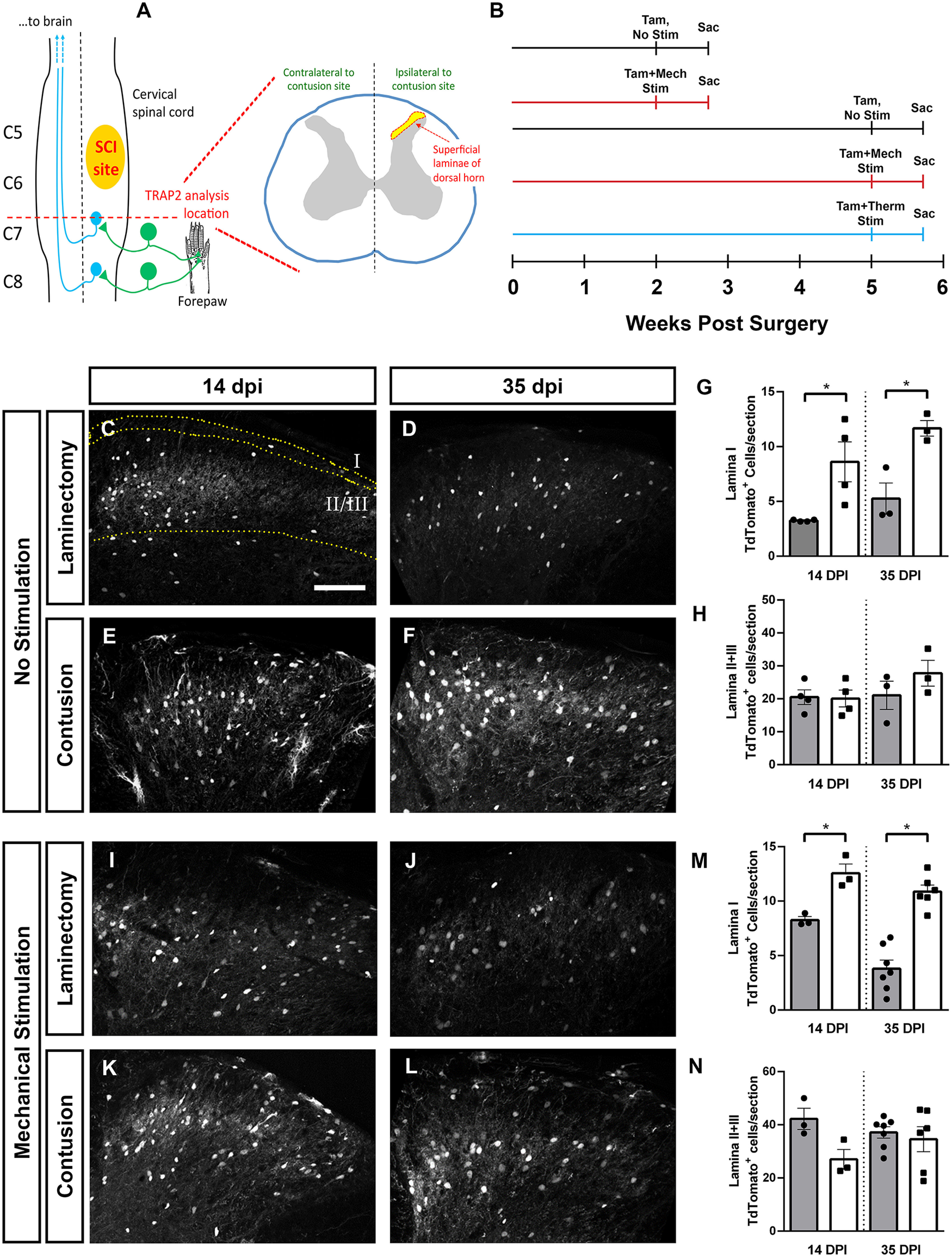
Figure 2.

Cervical SCI altered DH neuron activation in a lamina-specific manner. Unilateral C5/6 contusion or C5/6 laminectomy-only control was delivered to the right side of FosTRAP2 mice, and TdTomoto+ cell counts were performed in the intact, ipsilateral DH at C7/8. Diagram of injury and site of analysis (A). Activated neurons were quantified at 14 and 35 dpi, in the absence of ipsilateral forepaw stimulation, following mechanical stimulation of the ipsilateral forepaw (at 14 or 35 dpi), or following thermal stimulation of the ipsilateral forepaw (at 35 dpi). Timeline of experiments (B). Representative images of the DH at 14 d (C) and 35 d (D) after laminectomy, and 14 d (E) and 35 d (F) post-SCI. Quantification of Lamina I activated cell counts (G), and Laminae II+III activated cell counts (H). Representative images of DH following ipsilateral forepaw stimulation in laminectomy animals at 14 d (I) and 35 d (J), and contusion animals at 14 d (K) and 35 d (L). Quantification of activated neurons in Lamina I (M) and in Laminae II+III (N). Gray bars in graphs: laminectomy-only; white bars in graphs: cervical contusion. Scale bar: 100 µm.
