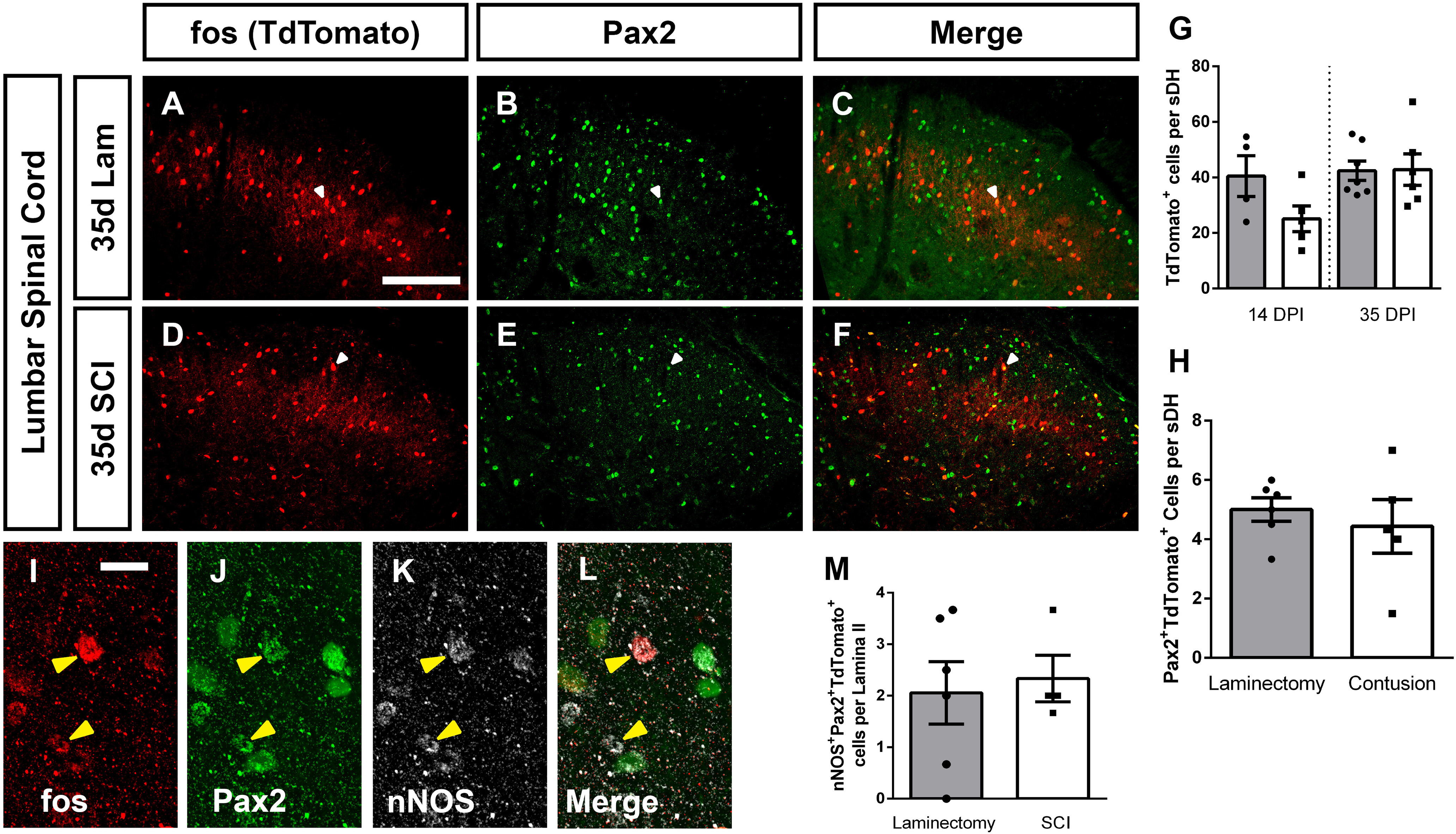
Figure 8.

Cervical SCI did not alter DH neuron activation in lumbar spinal cord. Representative DH images from L4/L5 spinal cord 35 d after laminectomy: TdTomato (A), Pax2 (B), merge (C), 35 d postcontusion: TdTomato (D), Pax2 (E), merge (F). Quantification of the number of TdTomato+ (activated) cells per superficial DH section (G), and of Tdtomato+Pax2+ cells per superficial DH (H). High-magnification representative images of TdTomato+nNOS+Pax2+ triple-labeled cells: TdTomato (I), Pax2 (J), nNOS (K), merge (L). Quantification of triple-labeled cells (M). Gray bars in graphs: laminectomy-only; white bars in graphs: cervical contusion. White arrowheads indicate double-labeled cells (in panels A–F); yellow arrowheads indicate triple-labeled cells (in panels I–L). Scale bar: 100 µm.
