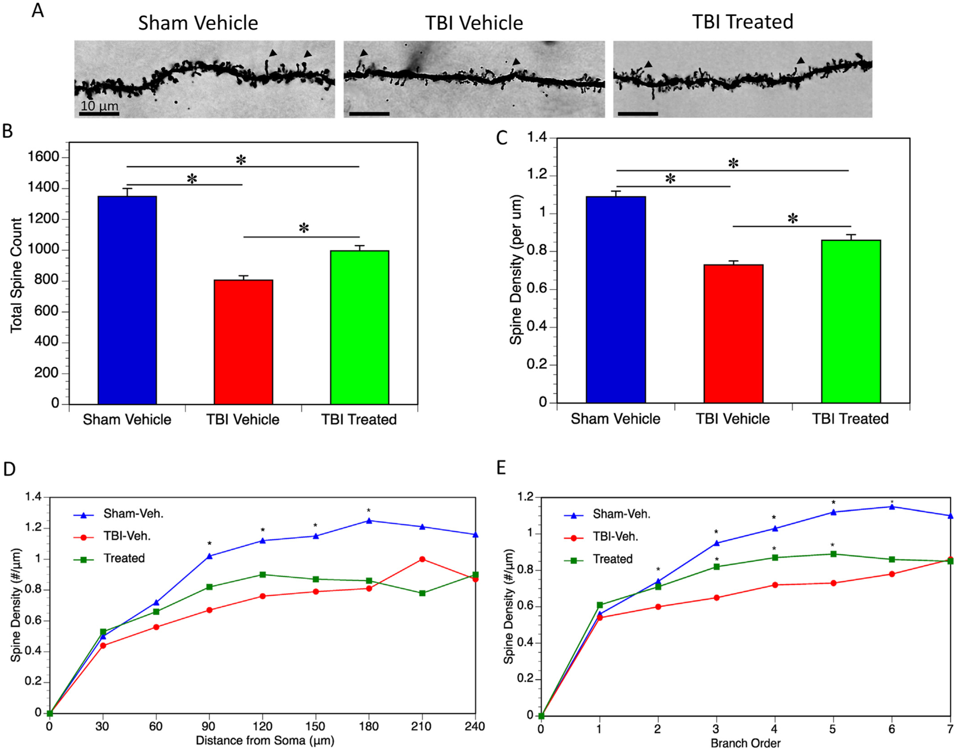
Fig. 3.

Dendritic Spines. (A) Dendritic spines of Golgi stained neurons of the dentate gyrus of the hippocampus were analyzed. Arrows indicate examples of dendritic spines. (B) Total spines among the 5 neurons analyzed per mouse was quantified. Total spines were significantly reduced in TBI vehicle mice when compared to sham (p < 0.0001). Total spines were increased in TBI treated mice compared to TBI vehicle mice (p = 0.0021), however there was still a significant reduction in total spines when compared to sham (p < 0.0001). (C) The same trend was observed in spine density (per μm). Both TBI groups showed a reduction in spine density compared to sham (both p < 0.0001), while TBI treated mice did have significantly higher spine densities compared to TBI vehicle mice (p = 0.0016). (D) Spine density was quantified based on distance from the soma. TBI vehicle mice had significantly reduced spine density 90–180 μm from the soma when compared to sham mice (p < 0.0001 for all distances). TBI treated mice had a slightly higher trend than TBI vehicle mice, however, this was not statistically significant. (E) Spine density was quantified based on branch order. TBI vehicle mice had significantly reduced spine density at orders 2 through 6 when compared to sham mice (p = 0.046 for the second branch order, p < 0.0001 for the 3rd – 6th branch orders). TBI treated mice showed some increase in spine density over TBI vehicle mice at the 3rd (p = 0.011), 4th (p = 0.029), and 5th (p = 0.018) branch orders.
