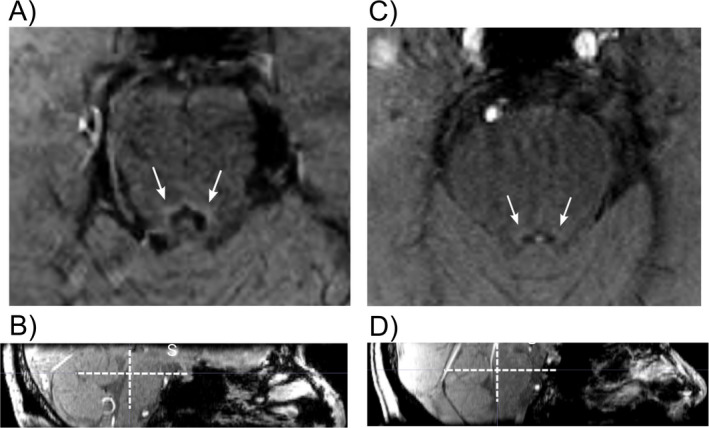
Figure 3.

Single slices from a 3‐Tesla magnetization transfer contrast MRI of the pons by our group. (A) (Axial) and (B) (sagittal) are from a 62‐year‐old healthy control, showing a healthy LC with clear contrast compared to the rest of the pons. (C) (Axial) is (D) (sagittal) are from 71‐year‐old patient with Alzheimer's disease with reduced contrast at the site of the LC, showing evidence of degeneration. White arrows and crosses highlight locus coeruleus (LC).
