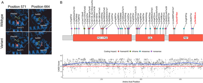
Figure 1.

Calpain‐1 structure with novel variants, variant distribution, and CADD PHRED scores. (A) The sites of predicted amino acid exchange in the PEF domain in the proband compared to wildtype. Mutated amino acids are colored in light red, interaction partners in light green, and hydrogen bonds in yellow. (B, upper panel) Schematic of the calpain‐1 primary protein structure. Disease‐associated variants identified in the literature are annotated along the protein with colored dots representing coding impacts. Novel variants identified in this report are labeled in red. (B, lower panel) CADD PHRED scores for all possible missense variants aligned to the CAPN1 protein structure. The recommended cut‐off for deleteriousness (20) is depicted as a red line. C2L, C2‐like domain; PC, protease core domains; PEF, large subunit of the penta‐EF‐hand domains. [Colour figure can be viewed at wileyonlinelibrary.com]
