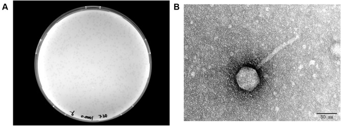
Figure 1.
(A) Morphology of IME268 plaques. Phages were plated in LB agar and overlaid with a liquid culture of K. pneumoniae 1733. The plates were incubated at 37°C. Clear, well-defined IME268 plaques were observed and photographed. (B) The morphology of phage IME268. IME268 was negatively stained with 2% phosphotungstic acid (PTA) and examined by transmission electron microscopy (TEM) at an accelerating voltage of 80 kV. The scale bar represents 50 nm.

