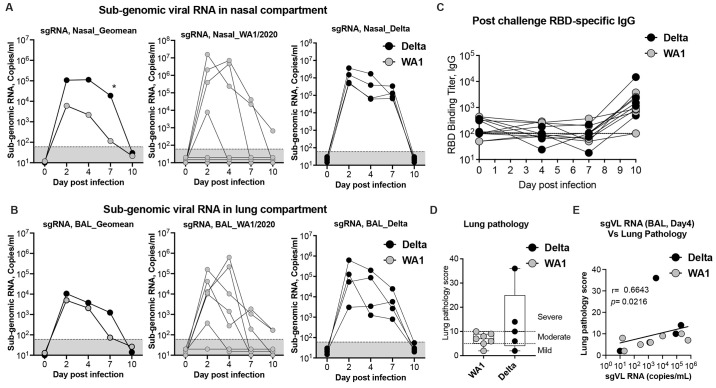
Fig. 5. Viral replication kinetics of WA-1/2020 and delta (B.1.617.2) after intranasal (IN) and intratracheal (IT) infection in unvaccinated rhesus macaques.
(A-B) Viral replication kinetics in the nasopharynx of the upper respiratory airways (upper panel) (A), and in bronchoalveolar lavage (BAL) fluid from the lower respiratory airways (lower panel) (B) in unvaccinated rhesus macaques following intranasal (IN) and intratracheal (IT) infection with WA-1/2020 (grey), and delta (B.1.617.2) (black) isolates. Replication was monitored from infection on day 0 until day 10 post-infection. Geometric mean titers (GMT) of sub-genomic RNA (sgRNA) copies/ml in log10 virus RNA copies per ml in nasopharyngeal and BAL samples (A), and from individual animal are shown. The limit of detection (<67 sgRNA copies/ml) is indicated in black dotted line. (C) RBD (WA-1/2020)-specific serum IgG responses in post-challenge serum. Each sample was tested in duplicates. (D) Lung pathology score at day 10 post-infection. (E) Correlation analysis between lung pathology scores and subgenomic viral loads (copies/mL). The Spearman rank test was used to perform correlation analysis. Each circle represents one animal. Comparison between groups was determined using a Mann–Whitney test. Error bars indicate the interquartile range with the median indicated by a horizontal line. Bars and columns show mean responses in each group ± SEM; Mann–Whitney test: ∗p < 0.05, and **p < 0.01. Dotted lines in viral loads reflect the limit of detection.

