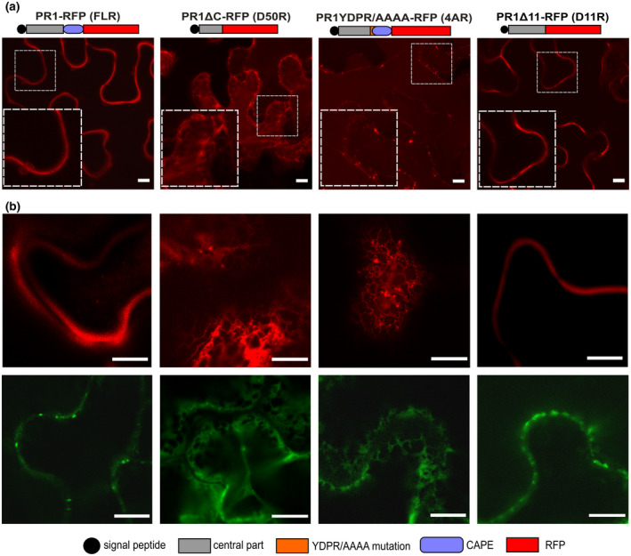
FIGURE 4.

Subcellular localizations of red fluorescent protein (RFP)‐tagged PR1 variants. (a) Schematic representation of RFP‐tagged PR1 variants and their localization in infiltrated Nicotiana benthamiana leaves. Insets depict a magnified view of the signal in the dashed squares. A strong extracellular signal for FL and D11 variants, and endoplasmic reticulum and vacuolar localization of D50 and 4A was observed. In all cases a minor portion of the vesicular signal was detected as well. Bars = 10 μm. (b) The representative images showing corresponding localizations of FL, D50, 4A, and D11 variants tagged with RFP (upper row) and GFP (lower row). Bars = 100 μm
