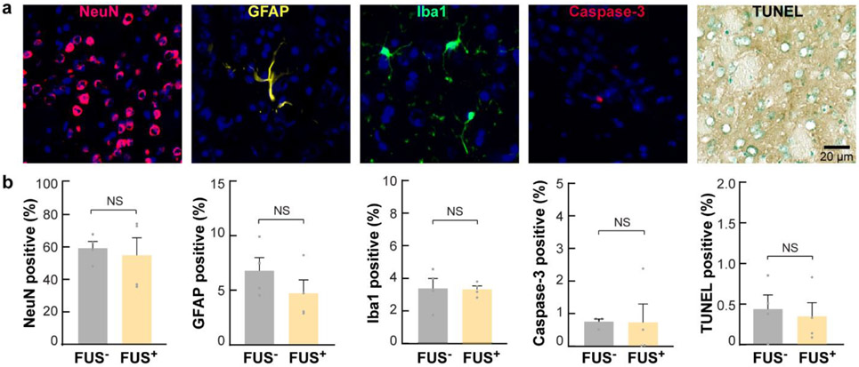
Fig. 6. Sonothermogenetics is safe at the cellular level.
(a) Evaluation of neuronal integrity, inflammation, and apoptosis after FUS exposure in the FUS-targeted brain location using immunohistochemical staining of neurons (NeuN), astrocytes (GFAP), microglia (iba1), caspase-3, and TUNEL. In the fluorescence images, blue indicates the DAPI-stained nuclei, and other pseudocolors indicate different cell types. (b) Percentages of positively stained cells to DAPI-stained cells within the FUS-targeted brain location in FUS-sonicated mice compared to those in mice without FUS sonication.

