Figure 3.

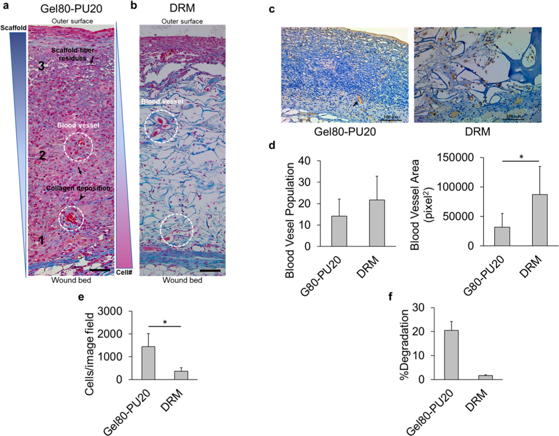
(a) Trichrome staining of acellular electrospun Gel80–PU20 membranes on the mice after 20 days, illustrating cell infiltration from the wound bed (zone 1) to the surface (zone 3) at the center of the wound (arrows: scaffold fiber residues; arrow head: collagen deposition; dashed circles: blood vessels.). The scale bar is 50 μm. (b) Trichrome staining of acellular DRM after 20 days on the mice. The scale bar is 100 μm. (c) CD31 staining of acellular electrospun Gel80–PU20 membrane and DRM on the mice after 20 days, showing blood vessels. (d) Quantification of blood vessel population and area on the image field, *P < 0.05. (e) Scaffold cellularity quantification per image field shown in (a,b). (f) Scaffold degradation after 20 days on the wound.
