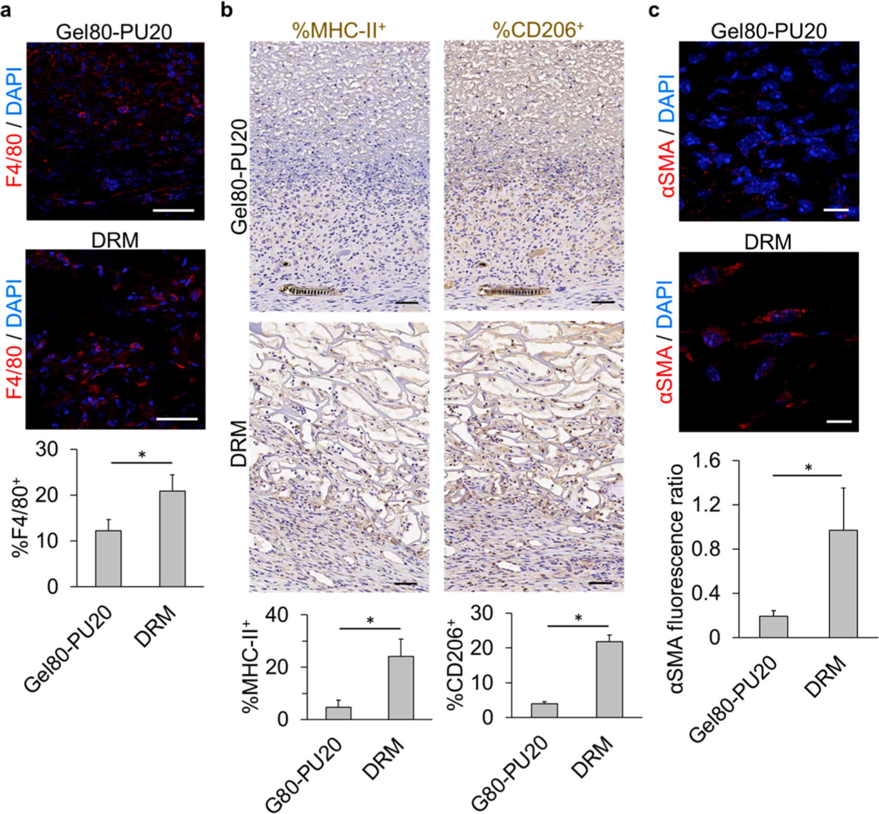
Figure 4.

Immunostaining of mouse wound sections covered by acellular electrospun Gel80–PU20 membrane and DRM after 20 days. (a) F4/80 staining for macrophages on the scaffolds and quantification of the F4/80+ cells (*P < 0.001). The scale bar is 50 μm. (b) Staining for MHC-II (M1) and CD206 (M2) and quantification of the stained cells (*P < 0.001). The scale bar is 50 μm. (c) Assessing the scaffolds for the presence of αSMA+ HDFs and quantification of the αSMA content by measuring the αSMA fluorescence ratio in each image field (*P < 0.001). The scale bar is 10 μm.
