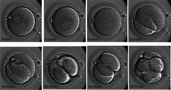
Fig. 1.
The sequential development of one of the embryos from the second patient’s last cycle which showed paternal contribution. At the beginning (0 h) and 5 h, a single pronucleus can be clearly seen as marked in a circle. By 8 h, the pronucleus was faded and cleavage division started around 10 h. Two and four cells were seen by 14 and 30 h, respectively. However, the embryo has reverse cleavage and became 3-cell around 40 h and reached to 8-cell by 60 h where it was biopsied

