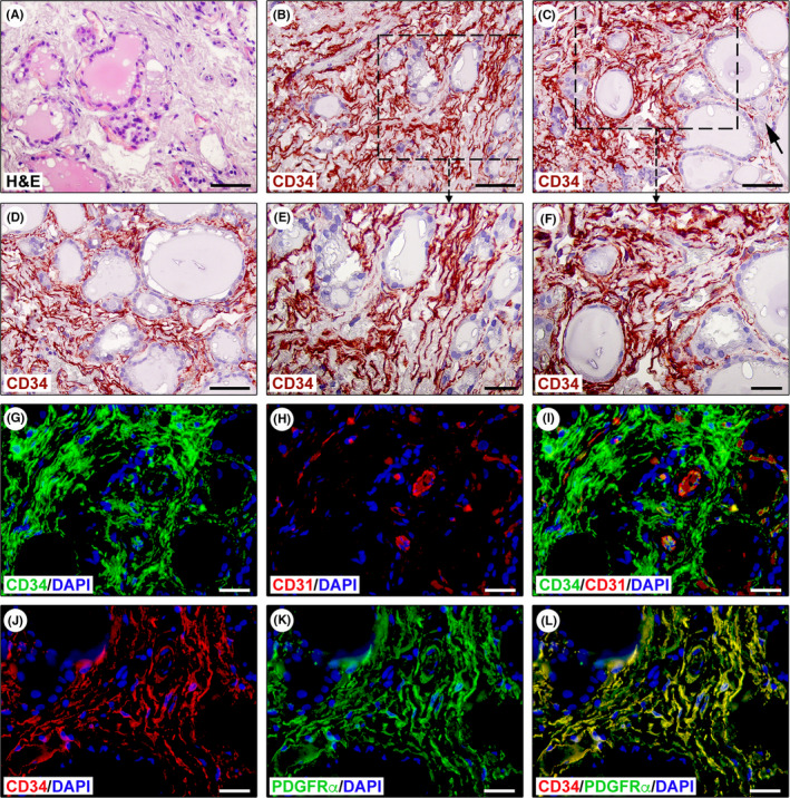
FIGURE 1.

Immunohistochemical identification of telocytes (TCs)/CD34+ stromal cells in normal human thyroid tissue sections. (A) Haematoxylin and eosin (H&E) staining testifying the normal appearance of thyroid tissue consisting of colloid‐containing follicles lined with epithelial cells (thyrocytes) and interfollicular reticular connective tissue harbouring fibroblasts, mononuclear cells, nerve fibres and small blood vessels and lymphatics. (B–F) CD34 immunohistochemistry with haematoxylin nuclear counterstain showing the presence of an intricate network of CD34+ stromal cells spread throughout the interfollicular connective tissue. Representative photomicrographs of tissue sections from three different specimens are shown. (E and F) Higher magnifications of the boxed areas in (B and C). CD34+ stromal cells exhibit the typical TC morphology, that is spindle‐shaped cells with very long cytoplasmic processes displaying an irregular calibre and a sinuous trajectory (E and F). CD34+ TCs are numerous around microfollicles, while they are less represented around macrofollicles (C, arrow). (G–I) Double immunofluorescence staining for CD34 (green) and pan‐endothelial marker CD31 (red) with DAPI (blue) counterstain for nuclei. The interfollicular network of TCs/CD34+ stromal cells lacks CD31 immunoreactivity and surrounds CD31+ microvessels (G–I). (J–L) Double immunofluorescence staining for CD34 (red) and platelet‐derived growth factor receptor α (PDGFRα, green) with DAPI (blue) counterstain for nuclei. All TCs/CD34+ stromal cells located in the interfollicular connective tissue display PDGFRα immunoreactivity (J–L). Scale bar: 50 μm (A–D), 25 μm (E–L)
