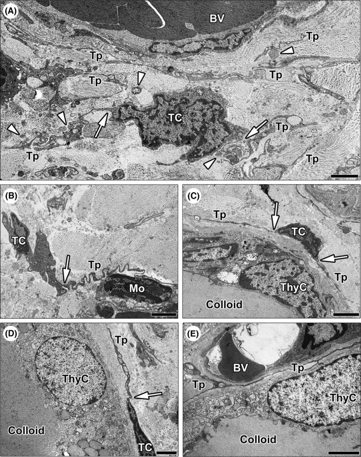
FIGURE 2.

Ultrastructural identification of telocytes (TCs) in normal human thyroid stromal tissue. (A–E) Representative transmission electron microscopy photomicrographs of thyroid ultrathin sections stained with UranyLess and bismuth subnitrate solutions. (A–D) TCs are ultrastructurally identifiable as stromal cells with long cytoplasmic projections (telopodes) characterized by a narrow emergence from the cell body (arrows) and a moniliform profile due to the alternation of thin segments (podomers) and expanded portions (podoms); the cell body of TCs is spindle‐shaped, oval, or piriform and mostly occupied by the nucleus. (A) Note a bipolar TC giving rise to two convoluted telopodes; the labyrinth‐like network formed by telopodes extends in between collagen bundles and around blood microvessels. Numerous extracellular vesicles are present nearby telopodes (arrowheads). (B) A TC projects a telopode with a very sinuous trajectory to intimately encircle and contact a mononuclear cell. (C and D) The telopodes of TCs are arranged around the basement membrane of thyrocytes lining colloid‐containing follicles. (E) Telopodes extend into the narrow interstitium between thyroid follicles and blood microvessels. Scale bar: 2 μm (A–E). BV, blood vessel; Mo, mononuclear cell; TC, telocyte; ThyC, thyrocyte; Tp, telopode
