
FIGURE 1.
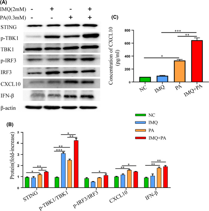
STING‐IRF3 pathway involvement in cellular model of diabetes and psoriasis. (A, B) Protein levels of STING, p‐TBK1/TBK1, p‐IRF3/IRF3, CXCL10 and IFN‐β were measured by Western blotting. (C) The expression level of CXCL10 in HaCaT cells was measured by ELISA. Data are represented as means ± SEM. *p < 0.05; **p < 0.01; ***p < 0.001; ****p < 0.0001