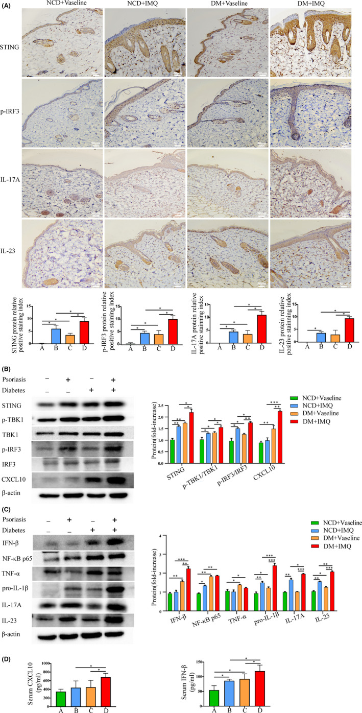
FIGURE 3.

Evaluation of STING‐IRF3 pathway in psoriasis and T2DM animal model. Group A, NCD + Vaseline, Group B, NCD + IMQ, Group C, DM + Vaseline, Group D, DM + IMQ. (A) The protein levels of STING, p‐IRF3, IL‐17A and IL‐23 in the skin tissue were measured by immunohistochemistry (×200). (B, C) The protein levels of STING, p‐TBK1/TBK1, p‐IRF3/IRF3 and CXCL10, and inflammatory cytokines IFN‐β, NF‐κB p65, TNF‐α, pro‐IL‐1β, IL‐17A and IL‐23 in skin tissue were measured by Western blotting. (D) Serum CXCL10 and IFN‐β of mice were measured by ELISA. Data are represented as means ± SEM (n = 6). *p < 0.05; **p < 0.01; ***p < 0.001; ****p < 0.0001; Scale bars, 50 µm
