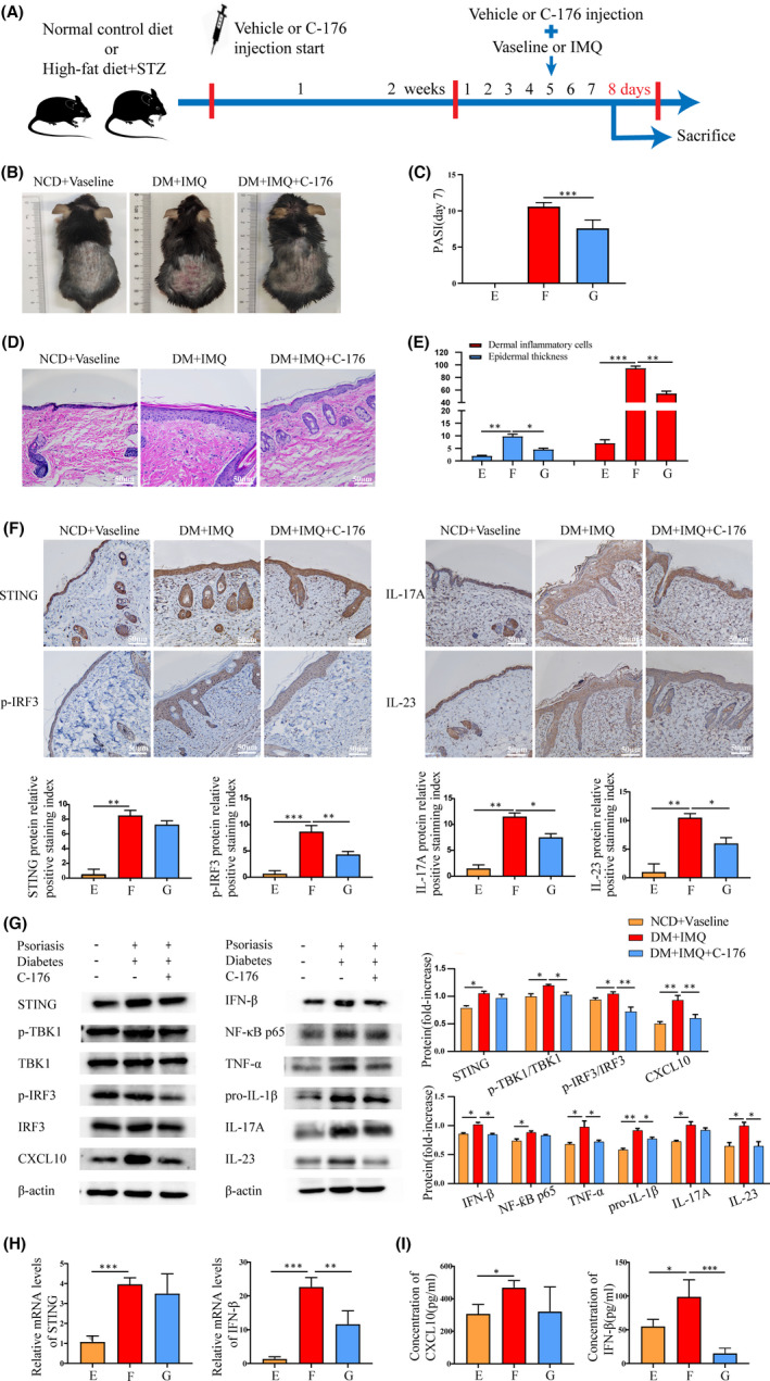
FIGURE 5.

Inhibition of STING‐IRF3 pathway in diabetic mice with psoriasis. Group E, NCD + Vaseline, Group F, DM + IMQ, Group G, DM + IMQ + C‐176. (A) Experimental procedure for the STING inhibitor: C‐176. After 7 days of intervention with IMQ, (B) visual changes in the skin on the back of each group. (C) Comparison of PASI scores of back skin of mice in each group. (D) Histopathological changes were observed under light microscope after HE staining (×200). (E) The epidermal thickness and the number of inflammatory cells in the dermis were analysed by Image‐Pro Plus 6.0 software (×200). (F) Protein levels of STING, p‐IRF3, IL‐17A and IL‐23 in skin tissue were measured by immunohistochemistry (×200). (G) Protein levels of STING, p‐TBK1/TBK1, p‐IRF3/IRF3 and CXCL10, and inflammatory cytokines IFN‐β, NF‐κB p65, TNF‐α, pro‐IL‐1β, IL‐17A and IL‐23 in skin tissue were measured by Western blotting. (H) Total mRNA levels of STING and IFN‐β in skin tissue were measured by qRT‐PCR. (I) Serum CXCL10 and IFN‐β of mice were measured by ELISA. Data are represented as means ± SEM (n = 6 mice in each group). *p < 0.05; **p < 0.01; ***p < 0.001; ****p < 0.0001. Scale bars, 50 µm
