
Fig. 2.
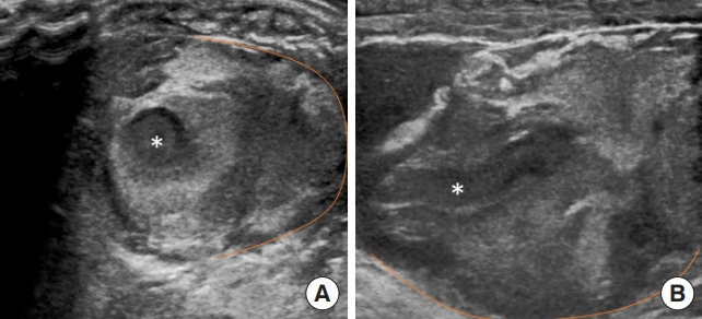
(A) Short axis view of first dorsal metacarpal artery (asterisk) with thrombosed material within lumen. Active extravasation of blood to the right of vessel with line indicating extent of hematoma within soft tissue. (B) Long axis view of first dorsal metacarpal artery showing hyperechoic thrombus within lumen (asterisk). Hematoma from extravasation of blood shown with line. The patient provided verbal informed consent for the publication of the research details and clinical images.