Fig. 1.
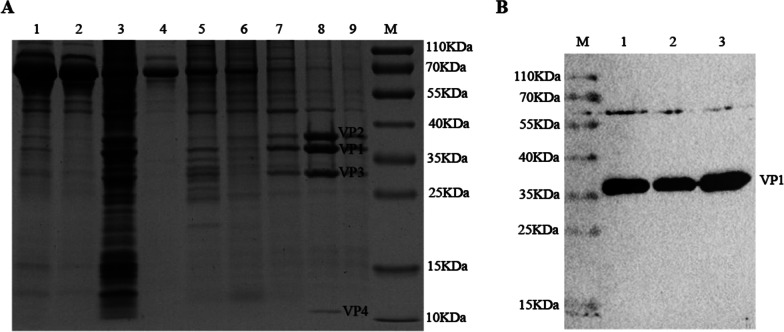
Immunogen preparation. A Components of the samples during ultra-high speed centrifugation were analyzed by SDS-PAGE and visualized by Coomassie blue staining. 1: Clear cell debris; 2: Concentrated supernatant; 3: Concentrated precipitate; 4: Sample layer; 5: 20% sucrose layer; 6: 35% sucrose layer; 7: 50% sucrose layer; 8: Virus Bands layer; 9: 65% sucrose layer; M: Protein Marker. B After the virus was desucrosed, the rabbit-derived antibody against SVV VP1 protein prepared in our laboratory was used as a primary antibody, and the purified SVV was identified by western blot. M: Protein Marker

