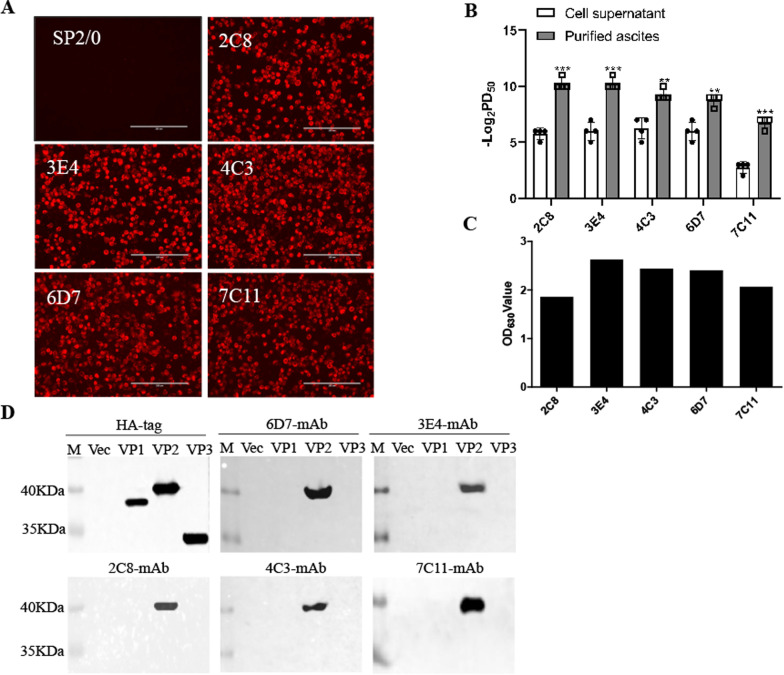
Fig. 2.
Functional analysis of monoclonal antibodies. A BHK-21 cells were infected with 0.01 MOI SVV-LNSY01-2017 for 36 h and then conducted for immunostaining analysis with 5 monoclonal antibodies. The mouse SP2/0 cell culture supernatant was used as a negative control. The cells were analyzed by an inverted fluorescence microscope. Scale bar: 200 μm. B The hybridoma cell supernatant and purified ascites of the five monoclonal antibodies were twofold multiple dilution and mixed with 200 TCID50 SVV. BHK-21 cells were incubated with virus-antibody mixtures to determine their neutralizing titers. C The prepared five monoclonal antibodies were used as primary antibodies in an indirect ELISA to test their reactivity with the virus. D 293 T cells were transfected with plasmids encoding VP1, VP2, or VP3. The cell lysates were conducted for western blot analysis with 5 monoclonal antibodies

