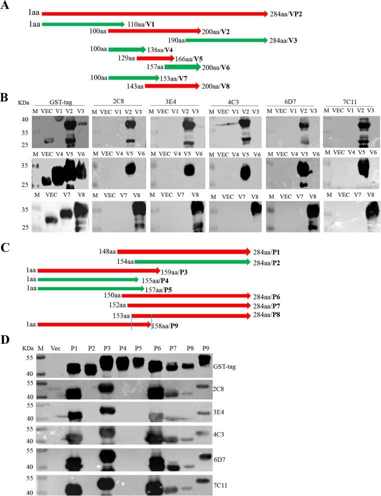
Fig. 3.
Identification of VP2 protein neutralizing linear epitopes by the antibodies. A Schematic diagram of the SVV VP2 protein fragment used for epitope mapping. V1: 1-110aa; V2: 100-200aa; V3: 190-284aa; V4: 100-138aa; V5: 129-166aa; V6: 157-200aa; V7: 100-153aa; V8: 143-200aa. B 293 T cells were transfected with plasmids encoding VP2 truncations for 24 h. The cell lysates were conducted for western blot analysis with the antibodies. C Schematic diagram of the SVV VP2 protein fragment used for epitope mapping. P1: 148-284aa; P2: 154-284aa; P3: 1-159aa; P4: 1-155aa; P5: 1-157aa; P6: 150-284aa; P7: 152-284aa; P8: 153-284aa; P9: 1-158aa. D 293 T cells were transfected with plasmids encoding VP2 truncations for 24 h, and then the cell lysates were conducted for western blot analysis with the antibodies

