Figure 6.
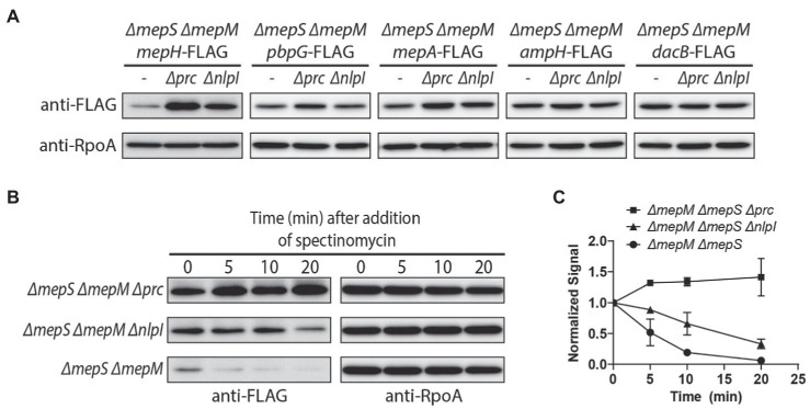
Prc and NlpI are involved in the negative regulation of MepH levels. (A) Comparison of DD-endopeptidase levels in the ΔmepS ΔmepM, ΔmepS ΔmepM Δprc, and ΔmepS ΔmepM ΔnlpI strains. The ΔmepS ΔmepM strains with DD-endopeptidase tagged with 3X FLAG and their Δprc and ΔnlpI derivatives were grown overnight in M9 glucose lacking casamino acids. The cells were washed and diluted in LB to an OD600 of 0.05. When the cultures reached an OD600 of 0.5, cells were harvested by centrifugation, resuspended in Laemmli buffer, and used for immunoblotting. (B,C) In vivo degradation assay of MepH-Flag. WJ309 (ΔmepS ΔmepM mepH-FLAG), WJ199 (ΔmepS ΔmepM Δprc mepH-FLAG), and WJ200 (ΔmepS ΔmepM ΔnlpI mepH-FLAG) were grown to an OD600 of 0.5 in LB, as described in (A). Spectinomycin was added to each culture to a final concentration of 500 μg/ml to block protein synthesis and aliquots were collected at the indicated time points for immunoblotting. The anti-FLAG signal of each sample was normalized to the anti-RpoA signal. Each experiment was performed in triplicates and the representative images are shown. (C) The normalized signal at the time of spectinomycin addition was set as 1 and the change in signal intensity at each time point was plotted for each strain. Error bars represent the standard deviation from triplicate measurements.

