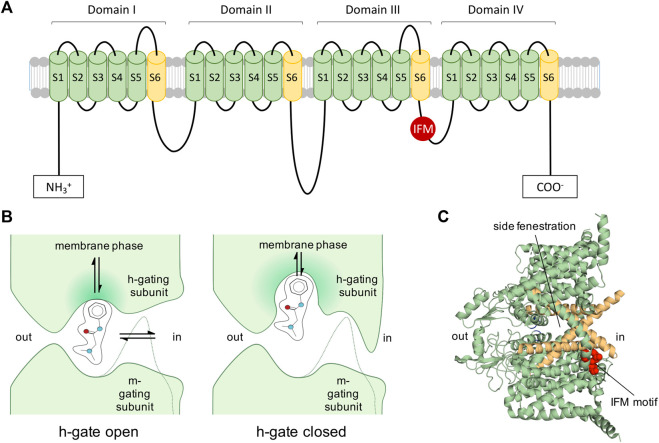
FIGURE 2.
Voltage-gated Navs as targets of LAs. (A): 2D representation of Nav. Inactivation motif is indicated by red circle labelled with IFM (isoleucine, phenylalanine, methionine). (B): View of 1977: Diagram of a LA molecule binding into the pore of a Nav indicating the activation gate (m-gate) and inactivation gate (h-gate) according to an early view and description of Nav modification (Hille, 1977). LAs were assumed to enter via the membrane phase or passing the m- and h-gate from the intracellular side. Many assumptions proved to be correct, such as the possibility for LAs to enter into the channel’s pore from the membrane phase. The figure was partially readapted from (Hille, 1977). (C): Cryo-EM structure of hNav1.7 (Shen et al., 2019) (PDB ID: 6J8G) depicting the side fenestrations, which allow LAs to enter the channel from the lipid phase and the inactivation motif (IFM-motif, shown in red), which was previously described as h-gate.

